

Introduction
Xylem and phloem are the types of plant vascular tissues. The primary function of both tissues is to assist the transport of water and mineral salts in the plant body. But these two tissues are different from each other. Xylem vs phloem microscopic differences are the primary topic discussed here.
A tissue is a group of cells that has a similar structure and is engaged in performing certain biological functions. Those types of tissue that cannot divide further are known as permanent tissue. Xylem and phloem are a type of complex permanent tissue.
Although these two tissues (xylem and phloem) are plant tissue, their structure and shape are completely different. The external structure of xylem and phloem can be seen with the naked eye. But their internal structure can be accurately reviewed with a microscope (2) & (4).
Microscopic definition of xylem
Xylem is a plant tissue and the internal structure can only be analyzed under a microscope. In 1858, Carl Nageli was first using the xylem word. A type of plant tissue that carries water and mineral salts to the plant body is called the xylem (1).
Features of xylem
The xylem tissues can be seen under a microscope when the plant is cut crosswise. Some features that are found in the analysis of the internal structure of xylem tissue by microscope are
1. The xylem cells have no nucleus.
2. There are two types of cells in the xylem known as parenchyma and fibers.
3. Its cell walls are thick and hard.
4. The cells which make up the xylem tube are dead that is, they do not contain cytoplasm.
5. Under a microscope the xylem tissue is star-shaped or small circles.
6. Xylem tissue color is green or blue-green under a microscope.
7. It is located in the center of the vascular bundles.
8. The cell wall of the xylem is made of mainly lignin and cellulose (1) & (4).
Xylem structure under a microscope
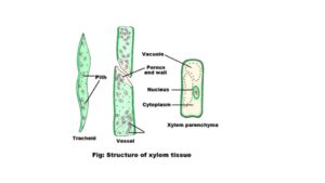
There are two primary vascular tissues of the plant, the xylem is one of them. The internal structure of the xylem can be described by the microscope as the inner structure can not be visible under naked eyes. Xylem tissue’s structure under a microscope is discussed below.
According to the basics of origin, there are two types of xylem tissue.
1. Primary xylem
It is formed during primary growth from procambium. This xylem is divided into protoxylem and metaxylem. Protoxylem is the first formed xylem. The lumen of its vessels is small. Metaxylem originates after the protoxylem but before the secondary xylem. It is found in all types of organs.
On the basis of the location of the protoxylem, the xylem is three types:
a. Endarch
Development of the xylem occurs from the center towards the periphery, that is, the protoxylem is present towards the center and the metaxylem towards the periphery in the stems. Such a type of xylem is known as endarch.
b. Exarch
Development of the xylem occurs from the periphery towards the center in roots, such type of xylem is exarch.
c. Mesarch
It is found in the ferns of some pteridophytes.
2. Secondary xylem
- It originated during secondary growth from vascular cambium.
- The secondary xylem is a cylinder-type cell.
- This tissue occurs towards the outer side of the primary xylem.
- Secondary xylem cells are made up of lignin.
- It is found mainly in stems and roots of only perennial dicots and gymnosperms.
- There are no special types of secondary xylem.
- It occurs towards the outer side of the primary xylem.
- Annual rings are present in the secondary xylem.
There are four types of cells that take part in the formation of the xylem.
a. Tracheids
Features
1. Tracheid cells are long.
2. It is a type of tubular cell.
3. These cells are located in the plants parallel to their longitudinal axis.
4. The ends of both sides of the tracheids are narrow and the width of the cell wall is curved or oblique.
5. Tracheids contain thin cell walls.
6. These cells transport water and mineral salts from the roots to the leaves.
7. They give rigidity to the plant.
8. Many times it stores water.
b. Vessels
The vessel is an elongated dead cell. It contains circular cross-sections. These cells are found in the xylem of flowering plants. Their cell walls are made of lignin. Its cells are usually thin and short. The primary function of a vessel is to transfer water from roots to every part of plants and to provide rigidity. These cells are less lignified. The length of the vessels is 10 cm. Vessel cells are only found in angiosperms.
c. Xylem parenchyma
Features
1. It is a part of complex permanent tissues.
2. These cells are living cells.
3. Cell walls of xylem parenchyma are made of cellulose.
4. Their main function is to store food.
5. These tissue cells help transport water and liquid food.
6. They have a nucleus and a protoplast.
7. The cells are colorless and there are big vacuoles.
8. Their cell wall is thick.
d. Xylem fibers
Xylem fibers are sclerenchyma cells. They are found in almost all dicotyledonous angiosperms. It is a non-living cell of the xylem. When these cells mature they lose their protoplast. Xylem fibers are located between the tracheids and vessels of the xylem. These cells also transfer water and liquid food to the plant body. The cell wall of xylem fibers is formed by lignin, so they provide rigidity to the plant (1) & (3).
Microscopic definition of phloem
It is a vascular tissue of a plant and was also discovered by Carl Nageli in 1858. There is a type of plant tissue that provides rigidity to the plant organs and also transports food and organic nutrients to the different parts of the plants. These types of plant tissue are called phloem tissue. This tissue is also known as bast or leptom.
Features of phloem
The internal structure of phloem tissue can only be analyzed under a microscope. A microscope is a device that can accurately analyze the internal structure of phloem tissue. Some features of phloem tissue that can be found by analyzing with a microscope are
1. The phloem is located in the external part of the xylem.
2. The cytoplasm is present in phloem tissue
3. It is a living tissue.
4. These tissues are formed from sieve tubes, companion cells, phloem parenchyma, and phloem fibers.
5. When phloem tissue is analyzed under a microscope, it is red or pink with toluidine blue.
6. Phloem’s cell walls are thin.
6. The phloem fibers are large and flexible.
7. Cytoplasm is present in phloem tissue cells.
8. These tissues carry food from the leaves to the growing parts and storage organs.
9. Under the microscope the phloem tissue is round or oval-shaped.
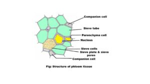
Phloem structure under a microscope
Like xylem tissue, Phloem is also a vascular tissue. The structure of phloem tissue is described accurately by electron microscopes. External structures of phloem tissues are visible to the naked eye. However, the internal structure is explained through a microscope.
Based on the origin and development, phloem tissue is divided into two parts.
1. Primary phloem
The primary phloem forms from the procambium during the primary growth of plants. It is found in all vascular plants. The tissue is formed in apical meristem tissue. Here sieve tubes are long and narrow. On the basis of development, the primary phloem is of two kinds.
a. Protophloem
This part is formed first. It is the outer part of the primary phloem. Protophloem is composed of narrow sieve tubes.
b. Metaphloem
Metaphloem is the inner part of phloem which is formed later. It is composed of broad sieve tubes.
2. Secondary phloem
Secondary phloem originates from the vascular cambium during secondary growth in plants. Here the sieve tubes are short and wide. The cambium cells by tangential divisions produce secondary phloem elements towards the outer side.
In most of the dicot stems, the primary phloem gets crushed and becomes inactive and the secondary phloem takes over the physiological activities. Cells are regularly arranged in the radial rows of the secondary phloem.
The phloem is the second vascular bundle tissue. When describing the structure of phloem tissue with an electron microscope, it is seen that this tissue is composed of three types of cells and one type of fiber.
a. Sieve tubes
Features
- They are living cells.
- Sieve cells are connected together to form sieve tubes.
- The cell wall of the sieve tubes is thin.
- Sieve tubes have protoplasm.
- Its cell wall contains lignin.
- Mature sieve cells have no nucleus.
- There are two types of sieve plates, simple sieve plates, and compound sieve plates.
b. Companion cells
Features
- It is a special type of parenchyma cells.
- The cells are thin and long.
- Protoplasm and nucleus are present in the companion cells.
- Each companion cell is closely combined with a sieve cell.
c. Phloem parenchyma
Features
- It is a type of parenchyma cell that is present in phloem tissue.
- These cells are cylindrical.
- The Phloem parenchyma has cytoplasm and nucleus.
- Cell walls are made of cellulose.
- Piths are present in the cell walls of phloem parenchyma.
d. Phloem fibers
Features
- These fibers are composed of sclerenchymatous cells.
- Phloem fibers are mainly present in secondary phloem.
- The fibers are elongated.
- They are dead cells.
- Phloem fibers have pits on their cell walls.
- It is long and has soft fibers (2) & (4).
Xylem vs phloem microscope differences
An electron microscope uses electrons (negatively charged atoms) instead of light to magnify an object. The wavelength of the electron is much shorter. Electrons can make tiny to very tiny things visible.
The xylem and phloem are the two main tissues of plants. Their internal structure is not simple. So the electron microscope is the only one that can accurately review the internal structure of these tissues of the plant (1). The differences between the xylem and phloem as described by the microscope are
Content |
Xylem |
Phloem |
| 1. Microscopic definition | Xylem is a type of plant tissue that carries water and mineral salts to the plant body. | A phloem is a type of plant tissue that transports food and organic nutrients to the different parts of the plants. |
| 2. Color | This tissue’s color is green or blue-green under a microscope. | Its color is red or pink with toluidine blue under a microscope. |
| 3. Shape | The xylem tissue is star-shaped or small circles. | Under the microscope, the phloem tissue is round or oval-shaped. |
| 4. Presence of cytoplasm | These tissues do not contain cytoplasm. | Cytoplasm is present in phloem tissue. |
| 5. Cell wall type | The cell wall of xylem tissue is thick and hard. | The phloem’s cell walls are thin. |
| 6. Location | It is located in the center of the vascular bundles. | This tissue is located in the external part of the xylem. |
| 7. Presence of conducive cell | Xylem tissue has two types of conducive cells. These are xylem tracheid and xylem vessels. | Phloem tissue is formed by one type of conductive cells, such as sieve cells. |
| 8. Fibers type | There is a type of fiber in xylem tissue. Known as xylem fibers. The fiber is sturdy and small. | Phloem fibers are large, soft, and flexible. |
| 9. Location in the vascular bundle | Xylem tissue is occupied in the center of the vascular bundle. | It is located on the outer side of the vascular bundle. |
| 10. Compose of cell wall | The cell wall of the xylem is made of mainly lignin and cellulose. | Its cell wall is made of only cellulose. These cells do not contain lignin. |
| 11. Cell type | Xylem consists of dead cells. | It is a living tissue. |
| 12. Tyloses | Formed in this tissue. | Tyloses are not formed in phloem tissue (1) & (3). |
Written By: Manisha Bharati
Reference
1. B Agarwal and V. K. Agarwal. Unified Botany, B.Sc. second Year. Shiva Lal Agarwal & Company Publications, Indore. Chapter: differentiation of primary and secondary tissues and their roles. Page no- 29 to 32 & 90 to 94.