

Know in one minute about Nephrons
|
Introduction
The human body is a hub of metabolic activities. Excretion is a process by which metabolic wastes are eliminated from the body. Kidneys are the primary organs that excrete nitrogenous wastes. In this article, we will know more about the role of the kidney along with the detailed structure of the nephrons.
Functions of the Kidney in the Body
1. Osmoregulation
Osmoregulation helps in maintaining the balance of the salt or minerals and water present in the body regardless of environmental conditions. Thus, the kidney helps in regulating the osmotic balance of the body. It regulates the amount of water and salt contents constant in the body
2. Excretion of nitrogenous waste
Nitrogenous wastes are harmful byproducts formed after protein metabolism. This waste if not removed from the body will form ammonia which is a toxic chemical. The kidney helps in the excretion of nitrogenous wastes, regulation of electrolytes, and salt concentration in the blood.
3. Regulation of acid-base balance
Acid-base balance or pH regulation is very important for the normal metabolism of the body and cell function. The kidney thus, helps in the regulation of the normal acid-base balance of the body. It regulates the bicarbonate concentration in the body. One of the functions is to absorb the carbonate and produce new bicarbonate thus balancing the acid-base equilibrium of the body (2).
4. Regulation of blood pressure
The kidney helps in regulating blood pressure by controlling the fluid balance in the body.
Nephrons are the building blocks of the kidney
Definition
Nephrons are the structural and functional units of the kidney. Around 1.2 million units of nephrons are there in each kidney. It is also known as uriniferous tubules.
Functions
Urine formation
The process of urine formation takes place in three steps in the nephrons. These are as follows
- Ultrafiltration – Filtration of blood plasma under high pressure through the glomerulus into Bowman’s capsule.
- Tubular reabsorption- Reabsorption of useful substances like glucose, amino acids, sodium, other salts, and water from the ultrafiltrate into the blood.
- Tubular secretion- Harmful substances like urea, excess salts, and chemical residues from medicines are secreted by the tubular cells of the nephron into the forming urine.
Types of Nephrons
1. Cortical nephrons
These nephrons occur in the renal cortex which is the outer dark region of the kidney.
2. Juxtamedullary nephrons
These nephrons occur both in the cortex and the medulla region of the kidney. Medulla is the inner pale zone of the kidney and the cortical region close to the medulla is called a juxtamedullary region.
| S/N | Cortical Nephrons | Juxtamedullary Nephrons |
| 1. | These nephrons occur in the cortex region (outer red dark zone of the kidney) | These occur in both the cortex and medulla region of the kidney.
|
| 2. | It constitutes 85% of the total nephrons. | Constitutes 15% of the total nephrons. |
| 3. | Structure: Glomeruli are small with short loops of HENLE extending a short distance in the renal medulla before turning back to the cortex. | Structure: Glomeruli are large-sized and located at the junction of the renal cortex and medulla(juxtamedullary) with long loops of HENLE penetrating deep into the medulla before turning back to the cortex. |
| 4. | They are surrounded by peritubular capillaries. | They are surrounded by vasa recta. |
| 5. | The rate of glomerular filtration is slow. | Glomerular filtration is fast. |
| 6. | Helps in the excretion of waste products in dissolved form in the urine. | Helps in the counter-current mechanism.
|
| 7. | They are more coiled. | Are less coiled. |
Structure of the Nephrons
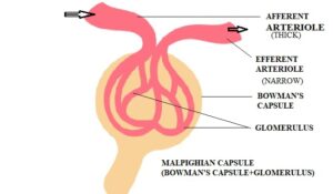
a. Renal corpuscle
It is the Initial filtering unit of the nephron. Also known as the Malpighian body.
-
Important structure
It consists of two parts namely
- Glomerulus- It is a mass of capillaries formed from the branch of the incoming renal artery (afferent arteriole thick) and again it unites to form a narrower efferent arteriole which joins the renal vein.
- Bowman’s capsule- It is a double-walled cup-like structure surrounding the glomerulus.
-
Function
Ultrafiltration (filtration of blood under high pressure) takes place in the glomerulus.
Most constituents of blood plasma can pass through except plasma proteins, colloids, and blood corpuscles.
-
Importance
Ultrafiltration gives rise to Glomerular filtrate which subsequently passes through the renal tubule to form urine.
-
Structure of Glomerulus
It is a tuft of capillaries formed at the junction of the afferent arteriole ( incoming) and efferent arteriole (outgoing ).
The endothelium of the glomerular capillaries has small holes called fenestra, which allow for plasma filtration and retention of plasma proteins and blood cells.
-
Structure of Bowman’s capsule
- Bowman’s capsule is a double-layered cup-shaped structure. The epithelium lining of the Bowman’s capsule is lined with podocytes (foot cells). They possess foot-like processes called pedicels. The space between pedicels is called slit pores through which glomerular filtrate filters.
- Outer parietal layer is composed of squamous cells and is continuous with the proximal tubule.
- The inner visceral layer surrounds the glomerulus.
- Basement membrane provides a bed for the capillary endothelium of the glomerulus and is mainly composed of a meshwork of collagen and proteoglycan fibrilae with large spaces through which fluid can filter.
b. Proximal convoluted tubule (PCT)
-
Functions
1. Reabsorbs sodium chloride, potassium, water, glucose, amino acids, bicarbonates, and amino acids.
2. Secretes ammonia and creatinine.
-
Importance
Maintains the pH and ionic balance of body fluids.
-
Structure
1. PCT is lined by cuboidal epithelial cells bearing brush border of microvilli to increase the surface area for absorption.
2. Tubule cells are rich in mitochondria which allow salt reabsorption by active transport
c. Loop of Henle ( Nephron Loop)
-
Functions
1. The descending limb is permeable to water and impermeable to electrolytes, water is reabsorbed from the filtrate making it hypertonic.
2. Ascending limb is impermeable to water but allows the transport of electrolytes from the filtrate making it hypotonic.
-
Importance
As the flow of filtrate in the two limbs of Loop of Henle is in opposite direction it forms a counter-current mechanism to concentrate urine and make it hypertonic.
- Structure
1. Descending limb of the loop of Henle has an upper thick part lined by cuboidal epithelium and has the same diameter as the PCT.
2. Mitochondria and microvilli are less in number than PCT.
3. The distal part of the descending limb is thin and lined by flat epithelial cells thinly scattered microvilli and very few mitochondria indicating a passive role in the ionic movements.
4. The thin segment of the ascending limb forms a major part of the Loop of Henle.
5. The thick segment of the ascending limb is lined by cuboidal epithelium cells with short epithelial microvilli and numerous mitochondria.
d. Distal convoluted tubule (DCT)
- Functions
1. Active reabsorption of sodium ions from the filtrate under the influence of aldosterone and water under the influence of antidiuretic hormone (ADH).
2. Tubular secretion of potassium, hydrogen ions, and other metabolic wastes like creatinine in the filtrate.
-
Structure
1. Thick ascending limb of Henle’s loop is continued as DCT.
2. It is lined by cuboidal epithelium.
Importance
The DCT maintains the water, salt, and pH balance of the blood.
Collecting Duct
-
Functions
1. Reabsorption of water takes place under the influence of ADH.
2. Aldosterone influences the reabsorption of Sodium.
3. This makes the filtrate hypertonic and is called urine.
-
Structure
1. Arises from the DCT and lined by cuboidal epithelium.
2. Passes through the renal cortex and medulla into the renal pelvis at the apex of medullary pyramids.
-
Importance
It plays a role in the maintenance of pH and ionic balance of blood by selective secretion of hydrogen and potassium ions and conserving body water.
The blood supply in nephrons
Afferent arteriole
- Renal artery branches to form a wider Afferent arterioles which break up into blood capillaries –Glomerulus.
- The blood leaves the glomerulus through a narrower Efferent arteriole.
- Efferent arteriole again forms a network of capillaries around the renal tubule to form the vasa recta.
Vasa Recta
- The efferent arteriole forms a network of capillaries around the renal tubule.
- The capillaries of this system unite to form the renal vein which joins the inferior vena cava.
Q&A
1. Where are nephrons located?
In the kidney.
2. How many nephrons are in each kidney?
About 1 .2 million
3. What are nephrons in the kidney?
Structural and functional units of the kidney.
4. What is the meaning of nephrons?
Greek word Nephros – meaning kidney.
5. Why are nephrons important?
Nephrons help in urine formation and the elimination of nitrogenous waste from the body.
Summary
- Nephrons are the functional unit of the kidney.
- Helps in the formation and elimination of nitrogenous waste.
- Excretion is the process of removing waste.
- They perform osmoregulation, and fluid homeostasis in the body.
- The renal corpuscle has a knot of blood capillaries called glomerulus in the depression of a cup-like structure –Bowman’s capsule.
- Glomerulus helps in ultrafiltration the first step in urine formation.
- The glomerular filtrate passes onto the next part of the renal tubule the Proximal convoluted tubule(PCT).
- Here tubular reabsorption of salts, water, and glucose takes place.
- Loop of Henle continues from the PCT and penetrates the renal medulla.
- The descending limb and the ascending limb form a hairpin-shaped loop and are responsible for concentrating the filtrate through the counter-current mechanism.
- The Distal convoluted region(DCT) arises from the ascending limb of the Henle’s loop
- Here reabsorption and tubular secretion also take place to maintain the pH of the blood.
- The collecting duct arises from DCT. Through reabsorption and tubular secretion, urine is formed here.
- Nephrons receive their blood through the afferent arteriole a branch of the renal artery.
- It breaks down into glomerulus and again reunites to form Efferent arteriole which leaves the Bowman’s capsule and forms a network of blood capillaries around the renal tubules called the vasa recta.
- These capillaries ultimately reunite to join the renal vein.
Written By: Ahana Mitra
References
Human physiology : The mechanism of body function – Vander , Sherman, Luciano.