

Earthworm circulatory system: A Quick Look
|
Earthworm circulatory system is of closed type structure. Blood flows in blood vessels and never comes in contact with tissues.
- Earthworms belong to the phylum Annelida.
- The body is metamerically segmented – externally by transverse grooves and internally by septa into many divisions.
- Each division is called a metameres.
- The earthworm body consists of about 100 to 120 segments or metameres.
- Seta the organ of locomotion is present in each segment.
- The first segment, the last segment and the clitellum lack seta.
The blood-vascular system of earthworms consists
- Blood
- Blood vessels
- Hearts
- Loops
- Capillaries
- Blood glands.
Blood
- The blood is red-coloured due to the presence of respiratory pigment haemoglobin.
- The haemoglobin remains dissolved in plasma in the form of erythrocruorin. It is a giant extracellular respiratory complex with 144 separate globin chains to carry oxygen.
- In the plasma is present many corpuscles. These corpuscles are amoeboid, nucleated and colourless. These corpuscles perform phagocytosis and thus protect the worm.
Blood Vessels
- Blood vessels are of two types – Collecting blood vessels and distributing blood vessels.
- Blood vessels are closed tubes with definite walls. These tubes break up into capillaries and branch into different parts of the body.
- The arrangement of blood vessels in the first thirteen segments is different from that behind the thirteenth segment.
Arrangement of blood vessels in the first 13 segments
Dorsal Longitudinal Blood Vessel
- Thick-walled and present on the mid-dorsal line of the alimentary canal.
- This blood vessel extends from the first segment to the last segment.
- It is the largest, distributing blood vessel. It is highly contractile. A pair of valves are present in each segment.
- The flow of blood in this vessel is from the posterior to anterior part of the body. The valves check the backflow of blood.
- It supplies blood to the first thirteen segments of the body.
- This vessel supplies blood to the buccal cavity, gizzard, oesophagus and pharyngeal nephridia.
Ventral Longitudinal Blood Vessel
- Thin-walled and present on the mid-ventral line of the alimentary canal.
- It is present from the second to the last segment of the body.
- It is non-contractile and valves are absent.
- Blood flows from the anterior to posterior part of the body. It distributes blood in these thirteen segments.
- It gives out branches called ventro tegumentary vessels one on each side. It supplies blood to the body wall and body organs of each segment.
Lateral Oesophageal Longitudinal Blood Vessel
- It is situated on the lateral sides of the alimentary canal. Present from the first to thirteen segments.
- Thin-walled, non contractile, valves are absent.
- Blood flows from the anterior to the posterior part of the body. Its main function is to collect blood from the body wall , reproductive organs, and nephridia.
Supra Oesophageal Blood Vessel
- It is the smallest blood vessel.
- Present from the 9th to 13th segment, on the dorsal side of the stomach.
- It collects blood from the stomach and gizzard.
- It receives blood from the lateral oesophageal vessels by two pairs of anterior loops.
Anterior loops
- Encircle the stomach in the 10th and 11th segments.
- It sends its collected blood by the latero –oesophageal hearts in segments 12 and 13 to the ventral vessel.
Ring Vessels
- These vessels are present in the wall of the gut. It connects the lateral oesophageal vessel to the supra oesophageal vessel. The blood flows upwards in these vessels.
Arrangement of Blood Vessels behind the 13th segment
Dorsal Longitudinal Blood Vessel
- Thick-walled and present on the mid-dorsal line of the alimentary canal. It has all the other features that it had in the anterior segments.
- It acts as a collecting vessel and in each segment, it receives :
- A pair of dorsointestinal vessel that bring blood from the intestine.
- A commissural vessel runs along the posterior border of each septum. It collects blood from the skin and nephridia.
Ventral Longitudinal Blood Vessel
- This runs along the mid-ventral line beneath the intestine and above the ventral nerve cord. It starts from the second segment and ends in the last segment of the body. It has thin walls without muscles and valves.
- It distributes the blood through the following :
- A pair of ventro tegumentary branches one on each side of each septum. It supplies blood to the inner body wall, integumentary nephridium, septal nephridia , gonads , seminal vesicle and spermathecae.
- A median ventro –intestinal branch that supplies blood to the floor of intestine. The branches in the intestine form blood plexus consisting of two networks in the intestinal wall.
Sub Neural Vessel
- It’s situated mid ventrally below the ventral nerve cord.
- It is a long, slender vessel extending from the 14th segment to the last segment.
- The direction of flow of blood is from the anterior to the posterior side of the body. It’s mainly a collecting vessel. In each segment, it collects the blood by a pair of ventral branches from the ventral part of the skin.
- In each segment, a pair of commissural vessels links the neural vessel to the dorsal vessel.
-
It collects blood from the ventral body wall and supplies some blood to the intestine.
Intestinal blood Plexus
- The intestinal wall of earthworms is traversed by an internal and external network of capillaries.
- The capillary network present at the outer surface of the intestine is known as the external plexus. It receives blood from the ventral vessel through ventro intestinal vessel and passes it on to the internal plexus.
- The capillary network present between the circular muscle layer of the intestine and the internal epithelial lining is the internal plexus. It absorbs the nutrients from the gut. It is connected to the dorsal blood vessel through the dorsointestinal vessels.
Commissural Vessels
- This connects the dorsal and the subneural blood vessels.
- It receives blood from nephridia, body walls and reproductive organs via capillaries. The blood is then sent to the dorsal blood vessel.
Integumentary vessels
- It originates from the ventral vessels.
- They supply blood to the integument for aeration.
- The aerated blood is collected by numerous capillaries of commissural vessel in each segment.
Nephridial Vessels
- These vessels originate from ventro tegumentary vessels of ventral vessel.
- It supplies blood to the nephridia.
Hearts
In each of the segments, the 7th, 9th,12th and 13th earthworms have four pairs of pulsatile, neurogenic hearts. The heartbeat originates in the nerve cells of the heart. Also termed as pseudo hearts or false hearts.
1. Lateral hearts
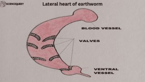
- A pair of hearts that are present on the 7th and 9th segments are lateral hearts.
- It connects the dorsal and ventral longitudinal blood vessels. Blood flows from above to below.
- Four pairs of valves are present here. Prevents the backflow of blood from ventral blood vessels to dorsal blood vessel
2. Latero-oesophageal hearts
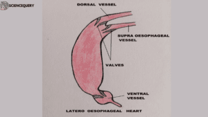
- The hearts of the 12th and 13th segments are called the lateral hearts.
- They connect the dorsal and the supra oesophageal blood vessels to the ventral blood vessels.
- Three pairs of valves are present to prevent backflow of blood.
Anterior Loops
- These are two pairs of loop-like vessels present in the 10th and 11th
- It connects lateral oesophageal vessels to the supraoesophageal vessels.
- Valves are absent in these vessels.
Capillaries
All the vessels in each organ pass into the network of the smallest capillaries. Blood from these capillaries flows into vessels across the body. From here blood flows down into the dorsal longitudinal blood vessel.
- The capillary network present at the outer surface of the intestine is known as the external plexus.
- The capillary network present between the circular muscle layer of the intestine and the internal epithelial lining is the internal plexus.
Blood glands
- Located in the 4th, 5th and 6th segments above the pharyngeal mass. These are several groups of small rounded, red colored follicles.
- Maintain blood volume and blood circulation.
- The walls have a syncitial wall enclosing a capsule containing a mass of loose cells.
- Blood glands are connected with pharyngeal nephridia and salivary glands.
- These glands help in the production of blood corpuscles and haemoglobin.
Circulation of blood in the body of Earthworm
From Dorsal longitudinal blood vessels
- Dorsal longitudinal blood vessels supply blood to the gizzard, which is collected by supra-oesophageal blood vessels.
- Also supplies blood to the buccal cavity, pharynx, and oesophagus. Blood from here is collected by lateral oesophageal longitudinal blood vessels.
- Supra oesophageal blood vessel receives blood from the lateral oesophageal blood vessels through anterior loops and ring vessels.
- From the supra oesophageal blood vessel blood is sent to the ventral longitudinal blood vessel through the lateral oesophageal heart.
- Dorsal longitudinal blood vessel also gives blood to ventral longitudinal blood vessels directly by lateral hearts.
- From the ventral longitudinal blood vessel arises the ventro tegumentary blood vessel in each segment.
- Ventrotegumentary blood vessel supplies blood to the anterior body wall, septa, nephridia and genital organs. This blood is collected by lateral oesophageal blood vessels. From here the blood passes to the supra oesophageal blood vessel via the anterior loop and ring vessels.
- From supra oesophageal blood vessels blood is again delivered to ventral longitudinal blood vessels via lateral oesophageal hearts.
- From the ventral longitudinal blood vessel arises ventro intestinal blood vessel from the 14th segment to the last segment.
- Ventro intestinal blood vessel supplies blood to the intestinal wall. From here the blood is collected by the dorso- intestinal blood vessel.
- Dorso- intestinal blood vessel sends this blood to the dorsal longitudinal blood vessel.
From Ventro tegumentary blood vessels
- Ventro tegumentary blood vessels are present in each segment. From the 14th segment to the last segment this blood vessel supplies blood to the nephridia, posterior body wall and other organs.
- From these organs, the blood is collected by the subneural blood vessels. From here the blood goes to the commissural blood vessel.
- A commissural blood vessel gives its blood to the dorsal longitudinal blood vessel. Subneural blood vessels and dorsal longitudinal blood vessels are connected by commissural blood vessels.
- From the commissural blood vessel arises a small blood vessel-septo intestinal blood vessel. This vessel supplies blood to intestinal walls. From here the blood is given to the dorsal longitudinal blood vessel through a dorso-intestinal blood vessel.
The function of the blood
- The atmospheric oxygen diffuses through the body wall into the capillaries. It combines with the haemoglobin to form oxyhaemoglobin. This oxyhaemoglobin is transmitted by the blood to the tissues.
- The oxyhaemoglobin breaks up to release oxygen to the tissues. This oxygen is used for oxidation of food stuffs to release energy.
- The blood distributes digested food to various body regions.
- Carbon dioxide diffuses into the blood and is carried in a dissolved state.
- It collects nitrogenous waste substances, and carbon dioxide and gives it to nephridia and the body wall.
Reference
Biology of earthworms by Edward, Clive A., et al.
| About the Author
Ahana Mitra, a zoology teacher with 22 years of experience, excels in teaching higher classes. Her strength lies in understanding students’ specific needs and simplifying complex biological concepts into clear, precise lessons. |