

Introduction
Do animal cells have a cell wall? well, the answer to this very curious question is “Animal cells do not possess a cell wall”. It is exclusively present in all plant cells, fungi, bacteria & cyanobacteria. Some protists also have cell walls.
The cell wall is basically the outermost, rigid & nonliving covering. In the year 1665, “Robert Hooke” first described it in the cork cells.
Animal cells have thin, delicate & living boundaries in comparison to plant cells. It is observed under the electron microscope and a clear bilayered lipo-proteinaceous membrane is seen, which is known as a cell or plasma membrane. The term was coined by Nageli. C & Cramer in 1855.
About cell walls in biology
In biology the outer covering of a cell is called the cell envelope, these are usually three types
- Cell walls present in plant cells & nonanimal cells
- Cell membranes are present in all types of animal cells.
- Glycocalyx is usually present in bacteria.
So by definition of the cell wall, we can say the outer thick and rigid permeable coat of a plant cell mainly formed of cellulose is known as the cell wall. It is a multi-layered structure.
Structure and function of cell wall
The cell wall is usually made up of pectin or hemicellulose. When we eat celery, the reason for that crunch we have is due to the cell wall.
All young fruits initially have a hardened appearance due to the above two components which gradually become disintegrated by an enzyme known as pectinase & that’s the time when we say the fruit has become ripe properly.
Composition of cell wall
The cell wall is composed of three layers. Namely from the outer to the inner side are the Middle lamella, Primary cell wall, Secondary cell wall, and occasionally tertiary cell wall.
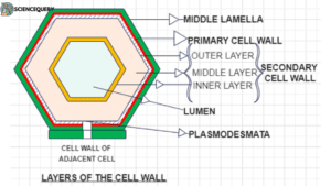
Middle lamella
- It is the outermost layer of the cell wall.
- Mainly made up of calcium and magnesium pectate.
- It is the nonliving layer that increases its volume by the accumulation of materials.
- It exists as a cementing layer between two primary cell walls of adjacent cells.
Primary cell wall
- Usually found in young plant cells and gradually disintegrates as the cell matures.
- This is capable of growth and it appears as a thin, elastic layer.
- Present inner to the middle lamella.
- Made up of 3% cellulose and 50% hemicellulose. Glycoproteins are also present.
- All meristematic and parenchymatous cells have only this cell wall.
Secondary cell wall
- This is present inner to the primary cell wall, just outside the plasma membrane.
- It can be differentiated again into outer, middle, and inner layers.
- This is the thickest layer.
- The composition of it is just opposite to the primary cell wall, here hemicellulose is 5 to 25% and 50 to 95% of cellulose is present.
- Other components like xylan, lignin, etc are there.
- Due to this more cellulose, this layer becomes rigid and nonelastic.
- Mainly found in collenchyma, sclerenchyma, and xylem vessels.
Tertiary cell wall
- Mainly seen in gymnosperms.
- Present inner to the secondary cell wall.
- Purely cellulosic in nature. Xylan is present in this layer.
Other structures associated with cell wall
Pits
Some areas of cell walls are not continuous, it lacks the secondary cell wall. These unthickened areas are known as pits.
They are complementary to the adjacent cell walls of another cell. Together they are called pit pairs. Two types of this pit present
- Simple pit: Where pit pores are uniform.
- Bordered pit: Pit pores are flask-shaped.
Plasmodesmata
These are the gaps between cell walls that connect cytoplasms of all adjacent cells. In this way, all the plant cells are connected to each other. Made up of desmo tubules which are extensions of smooth endoplasmic reticulum.
The function of the cell wall
- It gives structural integrity to the plant cells and also protects them from mechanical damage.
- Helps to stand erect, by providing mechanical support.
- It allows protoplasmic continuation by plasmodesmata. Which is important for the transport of substances.
- Prevents any osmotic bursting of cells.
- Helps in cell-to-cell adhesion and prevents the entry of undesirable molecules.
- Acts as protective covering barriers against pathogens.
Composition and role of the cell wall in different organisms
We can now understand that the cell wall is an additional layer of protection on top of the plasma membrane. You can find cell walls in both prokaryotes & eukaryotes and they are most common in plants, algae, fungi, and bacteria.
However, animals and protozoans do not have this type of structure. Cell walls tend to be rigid structures that help maintain the shape of the cell.
Table for differences between cell walls in different organisms
| PLANTS | ALGAE | BACTERIA | FUNGI | ||||
| Plant cell walls are primarily composed of cellulose, a complex carbohydrate polymer made up of glucose molecules.
Cellulose forms a rigid, strong, and fibrous network that provides structural support to plant cells. In addition to cellulose, plant cell walls may also contain other components such as hemicellulose, pectin, lignin, and proteins. |
The composition of algal cell walls varies depending on the type of algae. For example, diatoms have cell walls called frustules. Which are made of silica (silicon dioxide). Other algae may have cell walls made of cellulose, similar to plant cell walls, or a combination of different carbohydrates like agar, carrageenan, or alginates. |
|
Fungal cell walls are made up of a complex matrix of carbohydrates, proteins, and chitin
Chitin is a tough, nitrogen-containing polysaccharide that provides strength and rigidity to fungal cell walls. Unlike cellulose, which is found in plant cell walls, chitin is the primary structural component in fungal cell walls. |
Animal cell anatomy and plasma membrane
Animal cells differ from plant cells in many aspects. All of them are exclusively eukaryotic cells, i.e. they have a prominent membrane-bound nucleus and other membrane-bound organelles like mitochondria, etc.
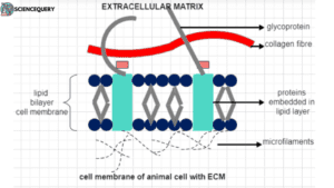
The cytoplasm of all animal cells is always covered by a thin, delicate, elastic & living boundary which is known as a cell or plasma membrane. Unlike plant cells here the rigid extra outer covering cell wall is not present.
Under an electron microscope, it seems two distinct layers are present here, of which one layer is the real membrane called plasmalemma or plasma membrane & the other is the extracellular matrix.
The chemical composition of this layer is protein 58%-59%, lipid 40%, and carbohydrate 1-2%.
Lipids
These are amphipathic molecules having a hydrophilic head (affinity to water) & two hydrophobic tails (repel to water). These form the continuous structural framework of the plasma membrane.
Proteins
These are known as membrane proteins.
Usually of two types- Integral membrane proteins- These proteins are completely or partially embedded into the membrane. for example, glycophorin & phorins.
Peripheral membrane protein- Present loosely at the outer surface of the membrane. for example, spectrin.
Carbohydrates
Oligosaccharides (sialic acid) are the main constituent.
To explain the structure of plasma membrane the best model we can name is the FLUID MOSAIC MODEL
According to this model, the plasma membrane is a continuous bilayer of phospholipid molecules in which globular proteins and sterols are embedded.
These arrangements resemble icebergs (protein) floating in the sea of water(phospholipids). These floating proteins give a false (quasi) fluid-like appearance, that’s why this model is also known as QUASIFLUID MODEL.
Some structural features of plasma membrane
- It’s about 7nm thick with the phospholipid layer as the basic layer.
- The hydrophilic heads of both layers face outward to the aqueous medium inside the cell & outside.
- Hydrophobic tails maintain a waterless environment inside the membrane
- Most floating proteins stay in the membrane because of hydrophobic amino acids which interact with the fatty acid hydrophobic tails to exclude water.
- Carbohydrate chains are attached to the proteins.
- In eukaryotes, cholesterol is also attached to the membrane but in prokaryotes, it is replaced by hopanoides pentacyclic sterols.
Cell membrane and outer barrier function of plasma membrane
Both the cell & plasma membrane are the name of the dynamic protein-lipid bilayer present at the outermost surface of the cell surrounding the cytoplasm & other cell organelles.
Apart from becoming the barrier between the inside and outside of the cell, it performs many other important functions-
Transport of the metabolites is the main function it provides. It can be divided into passive and active transport.
Passive transport is the transport of molecules along with the concentration gradient. From higher to lower levels of concentration.
This can be divided into two further ways-
Simple diffusion- the movement of the non-polar molecules from higher to lower concentration.
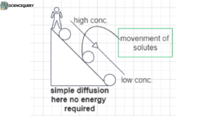
Facilitated diffusion- A special kind of diffusion where the movement of polar molecules across the membrane is facilitated by the formation of a metabolites carrier complex.
E.g.- entry of glucose in erythrocytes.
Active transport- Here molecules move against the concentration gradient. From lower conc. to higher. It requires energy in the form of ATP.
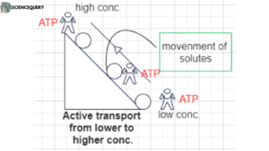
It can be further divided into two types-
Primary active transport- transport of Na+/K+ with the help of ATPase complex to maintain cellular volume.
Secondary active transport- Glucose transport in E.coli.
All the transportation at the cellular level is done by a plasma membrane.
Formation of blebbing-like formation of vesicles around the membrane.
Bulk transport also be done in the form of endocytosis-
Phagocytosis- engulfment of solid particles.
Pinocytosis- Intake of liquid particles.
Cell recognition & adhesion are also done by the plasma membrane with the help of carbohydrates as the site of cell recognition.
E.g.- macrophages recognize the carbohydrates present at the surface of worn-out rbc.
Plasma also works in the reception of different types of hormones & different types of secretions like polypeptide chains synthesized by ribosomes also occur.
Why is the cell wall absent in animals?
We know that the cell walls are rigid structures that give structural integrity & protective layer after the plasma membrane in plants. So we can assume that in the process of evolutionary changes animal cells get rid of this structure primarily to maintain its flexibility & locomotion. Animals need these two attributes for food, reproduction &, etc., which ultimately leads to the better use of the niche.
Niche is used to describe the multidimensional role an organism or population plays within its community or ecosystem.
Like tigers that live in a particular ecosystem, they are the alpha predator in that ecosystem where no one can harm them when they become adults, except humans.
Animals developed more unique and extensive cell signaling pathways, like we can see in our nervous system, immune system, etc. Which again makes them suitable & advanced. If we have a rigid cell wall around our cells it would hinder these communication pathways.
Animals also have more complex organ systems than plants. That’s why they need more flexibility in the shape & structure of cells. With a rigid cell wall, red blood cells or neurons can’t be shaped like they are now.
As animals can regulate their internal salt & water balance more actively, the absence of a cell wall gives them more efficiency in regulating the osmotic pressure.
It’s important to note that the absence of a cell wall in animals is a result of evolutionary adaptations that have allowed animals to thrive in their respective ecological niches. It helps them to relate to their environment more precisely.
Adaptation of animal cells without cell wall
The absence of the cell wall gives animal cells many adaptations by which they will fulfill all of their biological & physiological functions more easily.
1. Flexibility
Will be the prime example again, like Diapedesis, a unique phenomenon done by white blood cells where they can change their shapes to move one capillary to another to fight against foreign intruders like amoeba, That’s only possible because WBCs don’t have any rigid structure. The same happens in the case of muscle contraction etc.
2. Extracellular matrix (ECM)
Animal cells have a well-built extracellular matrix (ECM) above the plasma membrane, which helps in cell migration, cell to cell adhesion. Most likely they help in wound healing, in the time of accumulation of cells.
3. Cell Junction
The absence of a cell wall gives rise to the formation of cell junctions to perform specialized functions like maintaining tissue integrity & cellular interactions. Desmosomes help the adhesion of cells in tissues while tight junctions create the cell-to-cell barrier.
- Many specialized cells in animals grow structures by which they can move, like cilia/flagella which helps in survival. We can name the locomotion of mucus in the respiratory tract or the movement of sperm.
- We need to remember cells in animals need to go through a series of stages during cell division. The absence of cell division actually helps to facilitate the process. In terms of change of shapes or getting the needed chemical signaling.
- Which ultimately give rise to new offsprings, growth & development, etc.
- In immune cells as macrophages, WBC can engulf the foreign elements more efficiently due to the absence of a cell wall and so can the cells during phagocytosis & pinocytosis.
- Transport of various materials is possible only by the presence of this semi-permeable dynamic cell membrane, not a rigid cell wall around it.
These adaptations collectively allow animal cells to perform a wide range of functions and contribute to the complex physiology and behavior of animals. The absence of a cell wall has enabled animals to evolve intricate systems for communication, movement, defense, and reproduction, ultimately contributing to their success.
Mechanism for structural support and protection in the animal cell
In the absence of a rigid cell wall, animal cells have evolved various mechanisms for structural support and protection. These mechanisms work together to maintain cell shape, integrity, and function.
Extracellular matrix
An extracellular matrix (ECM), is a dense complex network of proteins & carbohydrates. It provides structural support to the cell and anchoring of cells in tissue cells.
Collagen and elastin
Proteins like collagen and elastin contribute to the mechanical strength of tissues, while glycoproteins like fibronectin and laminin mediate cell adhesion and migration.
Specialized cell junctions
Animal cells are connected by specialized cell junctions that help maintain tissue integrity and facilitate communication between cells. These include tight junctions, gap junctions, desmosomes, etc.
The cytoskeleton
It is a dynamic network of protein filaments that provides structural support and helps maintain cell shape. It is made up of three components
1. Microfilaments (Actin Filaments): This provides mechanical support and plays a role in cell movement, shape changes, and cytokinesis (cell division).
2. Microtubules: They are involved in maintaining cell shape, intracellular transport, and cell division.
3. Intermediate Filaments: These provide mechanical strength to cells and tissues. Different types of intermediate filaments are found in different cell types.
Cellular organelles within animal cells, such as the nucleus and endoplasmic reticulum, contribute to structural organization and support specific cellular functions. For example, the nucleus contains genetic material and controls cellular activities, while the e.r plays a role in protein synthesis and lipid metabolism.
Collectively, all these mechanisms work in tune to provide animal cells with the necessary structural support and protection required for their diverse functions within the body.
Q&A
1. Why is there no cell wall in an animal cell?
The way animal cells evolved in the course of evolution, there is no need to have a rigid cell wall around it, it would hinder the cells to perform various functions which are possible now due to their flexibility
2. Do animal plants have a cell wall?
No, animal cells don’t possess any cell wall like plant cells.
3. Does an animal cell plant cell or both have a cell wall?
Plant cells have a rigid cell wall. None of the animal cells has a cell wall.
4. Do animal cells have a cell wall and or chloroplast?
Animal cells neither have a cell wall nor a chloroplast. Both of these things are the components of plant cells.
Summary
- Animal cells don’t have cell walls.
- In the process of evolutionary changes animal cells get rid of the cell walls, primarily to maintain its flexibility & locomotion.
- Only plant cells, fungi, bacteria & cyanobacteria have the cell wall.
- The composition of the cell walls in these different organisms is different. It’s a rigid, nonliving layer made up of an accumulation of carbohydrates.
- The main function of it is as a protective layer & gives structural integrity to the plant.
- The plasma membrane is present in both plant & animal cells as the outer layer which covers the cytoplasm.
- It’s a living layer made up of a phospholipid bilayer, with proteins embedded in it.
- Function as a transportation and protective layer.
- Animal cells develop many adaptations as they don’t have cell walls.