

Introduction
The thin membrane on the outside of the protoplasm of eukaryotic and prokaryotic cells is called the cell membrane. It is a major part of the cell that mainly helps in protecting different parts of the cell from external damage. The cell membrane is formed by the transformation of the outer part of the cytoplasm. Many scientists believe that this major part of the cell is formed from the endoplasmic reticulum. The word cell membrane was first used by C. Nageli and C. Cramer. It is made up mainly of proteins and lipids. However, there are differences of opinion regarding the structure of this cell membrane. The following is a discussion of different cell membrane models (1) & (4).
Cell membrane
The fine, elastic, living, and microscopic membrane outside the cytoplasm that maintains the shape of the cell is called the cell membrane. The cell membrane is also called the plasma membrane or plasmalemma. The cell wall is present outside the cell membrane of plant cells.
Cell membrane creates a barrier between the cellular and the external environment. Some animal cells have a soft and thin glycoprotein covering on the outside of the cell membrane called a cell coat or glycocalyx. The cell membrane of animal cells is composed mainly of mucopolysaccharidosis, glycolipids, and glycoproteins. Desmosome is formed by the connection of the cell membrane of two animal cells.
The cell membrane is not usually visible to the naked eye (except for the thin membrane in a boiled egg). Microscopic instruments are used to view the cell membrane. Various scientists have described the structure of the cell membrane in different ways. Below are the details of all these models (1) & (6).
Models of the cell membrane
There are two main models based on the description of the physical and biological properties of the cell membrane.
1. Bilayer model
A. Lipid membrane model
In 1902, scientists overtone proposed the lipid membrane model of cell membranes. Overtone presented a basic model for small solvent transport. The detailed concept of his model is described below (1).
B. Overtone’s model
Charles Ernest Overtone presented a very important model of the cell membrane. In the 19th century, scientists overtone noticed that the movement of molecules through membranes was related to their separation coefficient between water and oil. He described this model by observing the osmotic properties of the cell. According to Overtone, the cell membrane is composed of a thin layer of the membrane. This layer is called the mono-lipids layer.
He also noted that the absorption and transport of soluble substances in lipids occurs faster than in water-soluble substances. On the basis of this observation, he concludes that the cell membrane is composed of a single layer of lipids. This model is also called a mono-lipid theory. According to Overtone, cholesterol, lecithin, and fatty oil are the main components of lipids (2).
C. E. Gorter and F. Grendel model
In 1925, E. Gorter and F. Grendel, two scientists at Leiden University, were the first to say that the cell membrane is a bi-layered structure of lipids. That is, this part of the cell is made up of two lipid layers. According to them, the cell membrane is made up of two phospholipid layers.
Phospholipids are the main components of the cell membrane. This element is present in the cell membrane in the form of a bimolecular layer. One side of the phospholipids is on the outside of the cell and the other side is on the inside of the cell towards the cytoplasm. This model is also called the lipid leaflet model. The model developed on the basis of their experiments later became the basis of other models (3).
D. Sandwich model or PLP (protein-lipid-protein)
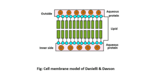
Discoverer
In 1935, Hugh Davson and James Danielli discovered this model of the cell membrane. Davson and Danielli proposed this model in terms of describing the molecular structure of the cell membrane.
Description of the model
According to Davson and Danielli, the cell membrane has a bi-layered phospholipid layer. These phospholipid layers contain protein layers on the outside and inside. According to them, there are two layers of lipid molecules with two edges on the outside of the cell membrane.
There are two layers of globular protein outside the lipid layers. This model is called the sandwich model because of the shape of the cell membrane. This is because the position of the protein layer on both sides and the lipid layer in the middle makes this model look like a sandwich.
This model states that the cell membrane has three layers. The structure of proteins and lipids is protein-lipid-lipid-protein. The total thickness of these three layers is about 80 to 100Å. There are also numerous vesicles on the outside and inside of the cell membrane. The thickness of these vesicles is about 5 to 10Å. The function of these vesicles is to transport small ions and water molecules (2) & (4).
2. Unit membrane model

The unit membrane model was first discovered by J. David Robertson in 1959. According to Robertson, all the membranes in the cell have the same basic structure. Therefore, known as “the unit membrane model”.
In this model, the structure of the cell membrane is to be trilamellar type that is, the cell membrane consists of two proteins and one lipid layer. According to him, the thickness of the cell membrane is about 75 Å. According to this model, the thickness of the protein layer in the cell membrane is about 20 Å, which is located on the outside and inside of the cell membrane. A layer of lipids is located between these two protein layers. The thickness of the lipid layers is about 35 Å.
The protein layer is aquatic and the lipid layer is water-resistant. In this model, the protein on the outside of the cell membrane is called the mucoprotein and the protein on the inside of the cell membrane is called the mucoid protein. Here the lipid layer is composed of two phospholipid layers. According to Robertson, not only the cell membrane but also the membranes of other organelles of the cell have the same structure (3) & (5).
A. Fluid mosaic model
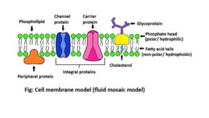
Discoverer
The model proposed by Singer and Nicholson to explain the structure of the cell membrane is known as the fluid mosaic model. In 1972, Singer and Nicholson discovered their fluid mosaic model. This model is now known as the universally accepted modern model.
Description of the model
According to Singer and Nicholson, the cell membrane is composed of phospholipids and protein molecules. These phospholipids are usually arranged in two layers. The phospholipid molecules in each layer have two sides. One side is called a hydrophilic head and the other side is called a hydrophobic tail. In the phospholipid layer, protein molecules are scattered. Besides, carbohydrates and other elements are scattered among the phospholipids.
Each phospholipid contains one molecule of glycerol and glycerol with two nonpolar fatty acid tails and one molecular phosphate head. Cholesterol molecules are present in the gaps between phospholipid molecules. There are many protein molecules floating between phospholipid and cholesterol molecules. There are two types of protein molecules located in the cell membrane based on the arrangement of molecules. These are-
- Peripheral proteins
These proteins are the outer or inner adjacent proteins of the phospholipid layer.
- Integral proteins
These proteins are partially or completely absorbed into the phospholipid layer.
Phospholipid molecules are constantly moving and vibrating. Then these phospholipid molecules move between the layers. Then the membrane looks like liquid or fluid. On the other hand, looking at the surface of the membrane, it can be seen that the protein molecules here look like mosaics.
That is why this model is called the fluid mosaic model. Phospholipid molecules and most protein molecules are associated with carbohydrate chains. These are called glycolipids and glycoproteins. Carbohydrate chains are always located outside the membrane (1) & (3).
B. Micellar or subunit model
- In 1963, micellar theory or subunit models were proposed by Hillier and Hoffman. According to them, the cell membrane is formed by the arrangement of globular subunits.
- According to this theory, the molecules of fatty acids present in the cell membrane are completely organized and bound in the water. This condition is called micelles.
- Each micelle contains layers of lipids. The hydrophilic edge of the lipid layer is present on the outside and the hydrophobic edge is present on the inside of the cell. This lipid layer contains a single layer of protein on both sides.
- The gap in the middle of the micelle is in the form of vesicles. The diameter of this vesicle is about 5Å. According to the subunit model, the cell membrane may be lamellar or micellar (1) & (4).
- Besides, In 1962 Fernandez-Moran and in 1963 Sjostrand discussed that the cell membrane consisted of globular units. These globular units are called elementary particles. The globules are about 30 to 60Å in diameter. And the globules are closely packed (2) & (3).
- Scientist Green also presented a model of the cell membrane. He described his model of the cell membrane in 1970. According to him, there are two types of the cell membrane. Such as monopartite membrane and multipartite membrane.
- An example of a monopartite membrane is the outer membrane of mitochondria. An example of multipartite is the inner membrane of the mitochondrial membrane (2) & (5).
Relationship between the bilayer model and micellar theory of cell membrane
There is a relationship between the bilayer model and the micellar theory of cell membranes. Scientific research supports both theories. The three layers of the unit membrane are clearly expressed by electron micrographs. The presence of subunits is known by chloroplasts and internal mitochondrial membrane.
Different membranes have different types of structures. That is, the transformation occurred between the by-layer model and micellar theory. For example, the membranes of chloroplasts and mitochondria appear to be constructed as a bi-layer backbone with a subunit or micellar structure on top (2) & (6).
Reference Books
1. B Agarwal and V. K. Agarwal. Unified Botany, B.Sc. Third Year. Shiva Lal Agarwal & Company Publications, Indore. Chapter: plasma membrane: bilayer lipid structure and functions. Page 217 to 221.
2. Ajoy Paul. Textbook for Zoology Honours (volume 1). Books and Allied (P) Ltd., Kolkata (India). Chapter: Plasma membrane. Page 314 to318.
3. Chandrasekhar Chakrabarti. A modern approach to a textbook of core Zoology, General & Honours. P Nirmala Library, A Publishing House under the Prestigious International Standard Book Number (ISBN) System. Kolkata, (India). Part – II. Chapter- cell and cellular organelles.