

Introduction
White blood cells and red blood cells are two important constituents of blood. Blood is the fluid connective tissue in the human body. It plays a major role in the transport of nutrients, oxygen, carbon dioxide, and other substances throughout the body. Blood comprises of two parts :
- Plasma
- Formed elements or blood corpuscles.
- Plasma is a light yellow-colored fluid mixture of water, proteins, inorganic salts, and other substances.
- The formed or cellular elements remain suspended in plasma. They are red and white blood cells and platelets. Red blood cells are the oxygen carriers in the body.
- White blood cells are of five types. Neutrophils, Eosinophils, Basophils, Lymphocytes, and monocytes. These help in preventing the body from infection and diseases.
- Platelets help in blood clotting.
Red Blood Cells
Are also known as erythrocytes. The human male has about 5.5 million RBCs / mm3. Females have 4.8 million RBC s/mm3.
1. Structure
- Red, flattened, biconcave, disc-shaped, about 8 microns ( 1 micron = 1 thousandth of a millimeter) in diameter. This shape provides a greater surface area across which the exchange of gases can occur.
- RBCs are flexible because they lack nuclei, mitochondria, endoplasmic reticulum, and other cell organelles. This helps them to travel through narrow spaces of capillaries.
- The plasma membrane has a network of proteins which allows for the flexibility of these cells.
- The cytoplasm of the RBCs is filled with hemoglobin (globin the protein part and iron-containing pigment heme). This gives blood its red color. Haemoglobin allows for the transportation of gases. A single erythrocyte can contain three million hemoglobin molecules.
- The plasma membrane of RBCs contains certain glycolipids which are known as antigens. These antigen accounts for ABO blood groups and Rh factors.
2. Life cycle
As the RBCs do not possess a nucleus they have to be renewed continuously. They cannot divide to produce new cells. After about 120 days of life span, they are replaced.
- RBCs arise from the stem cells in the bone marrow by a process called erythropoiesis.
- Hematopoietic stem cells differentiate into proerythroblasts.
- These proerythroblastic cells give rise to the early erythroblast stage.
- Ribosomes are synthesized which also synthesize hemoglobin. This stage is called the late erythroblastic stage.
- These erythroblasts develop into normoblasts.
- Normoblast develops into reticulocytes and loses its nucleus but it is not a RBC yet.
- Reticulocytes reside in the bone marrow for about three days before entering into circulation.
- Once in circulation, the reticulocytes mature into RBC or erythrocytes after one or two days.
- After 120 days they are destroyed in the liver, spleen, and bone marrow by the macrophages.
3. Functions
- Transport of oxygen – The erythrocytes are oxygen carriers. In the lungs, the hemoglobin binds with oxygen to form oxyhemoglobin. This is carried to the tissues for respiration and release of energy.
- Transport of carbon dioxide – RBC transports about 23% of CO2 from tissues into the lungs. Their hemoglobin forms carbamino–hemoglobin with carbon dioxide.
- Maintenance of pH of blood– Haemoglobin is a good acid-base buffer and is largely responsible for maintaining the pH of the blood. The acidity of blood reduces hemoglobin’s capacity to carry oxygen.
White blood cells
Also known as Leukocytes. Their number varies from 6000 to 9000 per mm3 of blood. These are the immune cells that circulate in the blood and lymphatic system.
Structure
- WBCs are larger than RBCs and lack hemoglobin. They are 12 to 20 micrometers in size.
- They have a nucleus, mitochondria, and other organelles.
- WBCs are of two types
1. Granulocytes
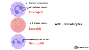
Contain granules and irregular multilobed nuclei in their cytoplasm. They are of three types :
a. Neutrophils
Most abundant WBC. About 60 – 65 % of WBCs are composed of this. They have a multilobed nucleus. They can be stained with both acidic and basic dyes.
b. Basophils
They constitute only 0.5 to 1% of total WBC. The nucleus is elongated and S-shaped. They can be stained with basic dye like methylene blue.
c. Eosinophils
They constitute 2 – 3% of the total WBC. They have a bilobed nucleus. Stain with acidic dye like eosin. Abundant coarse granules are seen in the cytoplasm.
2. Agranulocytes
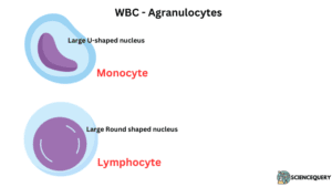
Granules are not found in the cytoplasm of these cells. They are of the following two types
a. Monocytes
Largest-sized WBC and has a kidney-shaped nucleus. Comprises 6-8% of total WBC. It is 12 -20 micrometers in size.
b. Lymphocytes
Small leucocytes with a large, round nucleus and little cytoplasm. They are 20 to 25% of total leucocytes. It is 6 to 14 micron metres in size. The lymphocytes produced in the bone marrow are B-lymphocytes. Those that are produced in the thymus are T-lymphocytes.
Life Cycle
The average life span of the WBCs is 12 to 20 days. The neutrophils live for a few hours.
- The bone marrow has pluripotential stem cells.
- These stem cells give rise to myeloid stem cells and lymphoid stem cells.
- Myeloid stem cells give rise to granulocytic WBC and monocytes.
- In the presence of Interleukin (IL) 3, 5 (chemical factors) and agranulocytic colony-stimulating factors( Ag-CSF), myeloid stem cells give rise to monoblast.
- Monoblasts differentiate into promonocytes and then into monocytes.
- These monocytes go to the bloodstream. Later they migrate to the tissues to become macrophages.
- Myeloid stem cells through another cell lineage division will give rise to myeloblast. IL 3, IL 5 and granulocyte stimulating factor (GSF) help in this division.
- These myeloblastes now differentiate into basophils, eosinophils, and neutrophils.
- Lymphoid stem cells divide into lymphoblast, in the presence of IL 3,5, Ag-CSF.
- The lymphoblast will now differentiate into prolymphocyte.
- Prolymphocyte now becomes the functional lymphocyte and leaves the bone marrow. It could be of two types: B-lymphocyte and T-lymphocyte.
- T-lymphocyte moves to another primary lymphoid organ Thymus and undergoes maturation. Then they become functional and released into the bloodstream.
Functions
Phagocytosis
On reaching the site of infection neutrophils and monocytes engulf microbes or damaged cells. This process is called phagocytosis. It protects the body against infections. Monocytes differentiate into macrophages which phagocytize dead and damaged tissue at the site of injury. This makes tissue repair easy. They secrete cytokines which modulate the immune response. Eosinophils contain hydrolytic enzymes, peroxidases that kill large parasites engulf bacterial toxins, and suppress allergy.
Diapedesis
Neutrophils show amoeboid movement. First WBC to arrive at the site of infection. They migrate towards the site of infection by squeezing out through the capillary walls to engulf and kill microbes. During an infection, neutrophils are produced more rapidly.
Inflammation and inflammatory reactions
The swelling caused at the site of injury is known as inflammation. Histamine is released by basophils during inflammation. Histamine is a vasodilator that causes dilation of blood vessels. So the capillaries become more permeable allowing tissue fluid, proteins, and white blood cells to accumulate in the damaged area.
Basophils produce heparin which prevents abnormal clotting of blood within the vessels.
Formation of antibodies
Lymphocytes produce antibodies in response to foreign antigens and destroy them. Both B and T lymphocytes provide memory for immunity.
Similarities between White blood cells and red blood cells
White blood cells |
Red blood cells |
| 1. WBCs are produced in the bone marrow from the single multipotent hematopoietic stem cell. | 1. RBCs are produced in the bone marrow from single multipotent hematopoietic stem cells. |
| 2. Worn-out WBCs are destroyed in the liver and spleen. | 2. At the end of the life span the RBCs are destroyed in the liver and spleen. |
| 3. WBCs are elastic. They can also squeeze themselves and sometimes even out of capillary walls. | 3. RBCs are elastic and can twist and squeeze as they pass through capillaries. |
| 4. WBCs have membrane surface receptors, proteinaceous in nature. This helps in recognizing threatening antigens in the body and destroys it by phagocytosis. | 4. RBCs have glycoproteins (antigens) on their plasma membrane that give the characteristic blood group to individuals. It also helps in detecting mismatched antibodies during blood transfusion. |
Differences between White blood cells and red blood cells
White blood cells |
Red blood cells |
| 1. WBCs are irregularly shaped with varying forms of nuclei. | 1. RBCs are biconcave disc-shaped and lack a nucleus. |
| 2. Five types of WBC are there: Eosinophil, Basophil, Neutrophil, Monocyte, and Lymphocyte. | 2. Only one type of RBC is found. |
| 3. Life span is 12 to 20 days. | 3. Life span is about 120 days |
| 4. Present in the cardiovascular and lymphatic system. | 4. Present in the cardiovascular system |
| 5. 6000 to 8000 WBC /mm3 of blood in both males and females. | 5. Males have 5 to 5.5 million RBC/mm3 and females have 4 to 4.5 million RBC /mm3. |
| 6. Colorless, presence of cell organelles and granules in the cytoplasm. It contains various chemicals like hydrolysates and peroxidases to help in the defense and immune system of the body. | 6. Contain respiratory pigment hemoglobin, red in color, lacks cell organelles. |
| 7. Produced in the bone marrow as well as lymph nodes, spleen, and thymus. Destroyed in the liver, spleen, and lymph nodes by phagocytosis. | 7. Produced in the bone marrow and destroyed in the liver and spleen. |
| 8. WBCs are mainly involved in the body’s defense mechanism through the production of antibodies, phagocytosis, and diapedesis. | 8. Transport oxygen from respiratory organs to all body parts. Carry carbon dioxide from all body parts to respiratory organs. Also carry hormones, nutrients, and other substances. |
| 9. WBCs constitute 1% of the total blood volume. | 9. RBCs constitute 25 to 50 % of the total blood volume. |
Q&A
1. What is the difference between red blood cells and white blood cells?
Red blood cells are red due to the presence of hemoglobin and non-nucleated, biconcave in shape. White blood cells are irregular in shape and possess nuclei. RBC helps in the transport of respiratory gases. White blood cells help in providing immunity to the body.
2. What is the relationship between red and white blood cells?
Red blood cells and white blood cells are formed from hematopoietic stem cells in the bone marrow through a process called hematopoiesis.
3. What happens if white blood cells are high?
If the white blood cell count is high it indicates an underlying infection or injury in the body. This condition is called Leukocytosis.
4. What happens if you have high white blood cells and low red blood cells?
High WBC causes Leukocytosis and low RBC causes anemia.
Summary
- White blood cells and red blood cells are constituents of blood the fluid connective tissue in the blood.
- Blood is composed of two parts, plasma the fluid part that remains suspended the blood corpuscles.
- Blood corpuscles are of three types – red blood cells, white blood cells, and platelets.
- Red blood cells (Erythrocytes) are a biconcave, disc-shaped, nonnucleated, and 7-micron meter in size.
- RBC is red in color due to the presence of iron-containing pigment hemoglobin (Hb). This can combine with oxygen and is delivered to all the parts of the body as oxygenated blood.
- RBCs also carry carbon dioxide from all parts of the body. The respiratory organs send it out of the body.
- In adult males, the number of RBCs is 5 to 5.5 million RBC/mm3. Adult females have 4 to 4.5 million RBC /mm3.
- RBCs are produced in the marrow of long bones by a process called erythropoiesis.
- The life span of RBC is about 120 days. After that, it is destroyed in the liver, spleen, and bone marrow.
- WBCs (Leucocytes) have a nucleus, colorless and irregularly shaped.
- Five types of WBC are there : (Granulocytes -Eosinophil, Basophil, Neutrophil),( Agranulocytes -Monocytes and Lymphocytes.
- WBC shows diapedesis and also helps in fighting infection, inflammation, and allergy in the body.
- Produced in the bone marrow (Leukopoiesis) as well as lymph nodes, spleen, and thymus. Destroyed in the liver, spleen, and lymph nodes by phagocytosis.
- WBCs also produce antibodies that kill or neutralize germs.