Introduction
A microscope is used to see objects that are not visible to the naked eye. The electron microscope is a breakthrough invention in microbiology. It is of two types based on its structure such as transmission electron microscope and Scanning electron microscope (SEM). This microscope accurately describes the smallest part of an object.
After the invention of the microscope, the structure of the object from 0.2 mm could be described using an optical microscope. But it was not possible to describe the structure of an object with a diameter less than 0.2 mm. Then in 1931, Ernest Ruska, a German physicist, invented the electron microscope. His discovery greatly improved science.
This device is widely used in microbiology, various experiments, medicine, laboratory, and many more. The main feature of this device is that it describes the magnified image of an object using electrons, the negative particles of matter, instead of light. Science the electrons are fast-moving particles, magnified images of objects can be obtained very quickly with the help of electron microscopes. The function that a simple microscope is unable to do can be done quickly by an electron microscope (1) & (2).
Scanning electron microscope
A scanning electron microscope (SEM) is a type of powerful electron microscope where objects are observed using fast-moving electron particles. This microscope observes a variety of organic and inorganic substances from 1 nanometer to micrometer.
In 1937 Manfred Von Ardenne first invented the scanning electron microscope. Although the scanning imaging method was first introduced in electron microscopes, it became known in biological and medical sciences many years later.
Before the discovery of this device in the history of science, the objects collected by electron microscopes did not have any 3D images. That problem was solved after the invention of this device. In 1942, the first scanning electron microscope was used in the United States to test the surface of solid objects (1).
Properties of Scanning Electron Microscope
- A scanning electron microscope (SEM) can accurately describe a 3D image of a sample.
- With the help of scanning electron microscopes, the image of the surface of the object can be captured perfectly.
- This device can magnify the image 10,000,000 times.
- In this device, the object is placed on a photographic plate and observed.
- Scanning electron microscopes makes it possible to take a large picture of the whole object.
- The resolution of scanning electron microscopes can be 1 nanometer or less.
- Samples obtained from the scanning electron microscope are white and black.
- A large number of samples can be analyzed simultaneously with a scanning electron microscope (3) & (5).
Process of scanning objects by SEM
The scanning electron microscope usually emits thermal electrons by heating the tungsten filament. It has a photographic plate behind the object. The reason for using tungsten in this microscope is that the melting point of tungsten is the highest and the vapor pressure is the lowest. Thus the thermal electrons that are generated are made of high energy.
A generated heat electron beam is emitted by a condenser lens with a diameter of 0.4 to 5 nanometer. When these initial electron beams act on the sample, they gradually lose energy. As a result of this interaction, scanning microscopes produce different types of symbols. These are secondary electrons, backscattered electrons, x-rays, sample flow, etc. Of all these symbols, the secondary electron is the most widely used. This is because the secondary electron can produce high-resolution images.
The symbols generated by the process are detected by a scanning electron microscope detector and a concept of the shape of the sample is obtained. On the back of this sample is a photographic plate. The function of this photographic plate is to capture the pattern of the magnetic field created by the electron particle hitting the object. As a result, a magnified image of the whole object is obtained from this microscope.
Parts of Scanning electron microscope (SEM)
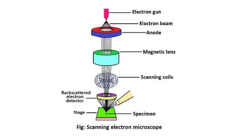
Scanning electron microscopes consists of several parts. These are electron guns, lenses, sample chambers, detectors, vacuum chambers, and scanning coils. The combination of all these parts allows a sample to be accurately observed and a picture of the whole object to be obtained from the sample. These parts of the scanning microscope are described below (2).
1. Electron gun
The electron gun of scanning electron microscopes transmits a large and stable amount of electricity to an electron beam. The electron gun is located at the top of the microscope and sometimes the electron gun is seen at the bottom of the microscope.
It is situated in the upper part of the electron column. This part is connected to the outside by a high voltage (30-40 kV) power cable. Electron guns act as a source of electrons. Its main function is to generate the electrons that are transmitted through the scanning electron microscope (1) & (2).
2. Lenses
The lens is used under a scanning electron microscope are not made of glass but made of magnets. The lens is used to focus the electron beam generated from the electron gun. Two or three condenser lenses are located under the electron gun of a scanning electron microscope. The main function of the scanning electron microscope lens is to help bend the flow of electrons (2) & (6).
3. Sample chamber
The sample chamber of a scanning electron microscope is the part where the sample is collected. In order to keep the sample stable, the sample chamber must be strong and separate. The sample chamber of SEM not only does the function of stabilizing the sample but also does other things like moving the sample from one place to another, placing the sample at different angles, etc. The sample stage and detector are located above the sample chamber (1) & (2).
4. Detectors
The scanning electron transmits electrons to all the objects in the microscope and observes the objects. The electrons in these objects are attracted by a device called a detector inside the electron microscope. And converts these electrons into electric currents. The main function of the detectors is to convert an electric current into an image and to present it on the screen (4).
5. Vacuum chamber
It is mandatory to have a vacuum chamber inside the scanning electron microscope. This is because electrons are scattered by any light element such as air. And in the case of deviation of electrons the exact concept of the object is not obtained from the sample. A vacuum pump is needed to remove air and other gases from the chamber to create a vacuum in the vacuum chamber (2).
6. Scanning coil
The main function of the scanning coil is to centralize the electron beam after it is emitted from the electron gun. After centralization, the scanning coils present in the electron microscope deflect this electron beam on two axes. This results in the sample being scanned (2) & (3).
There are some other parts of the scanning electron microscope that help observe the sample. These are power supply, a display device (computer), anode, sample stage, controllers, heater, and water chiller.
The scanning electron microscope has a positively charged plate called the anode. The anode attracts electrons and turns them into electron beams. The display device is the screen on which the image is displayed after the sample has been analyzed under a scanning electron microscope. The combination of all these parts accurately observes a sample and creates an image from the sample (2).
Principle of scanning electron microscope (SEM)
The scanning electron microscope (SEM) focuses on the primary electrons are emitted from the electron gun and many symbols are generated. These are secondary electrons, backscattered electrons, characteristic x-ray, sample flow, etc. The most used of these is the secondary electron, which is capable of producing high-resolution images.
These symbols are generated by the collision of the electron beam with the atom near the sample chamber. With the help of the secondary electron signal of a scanning electron microscope, it is possible to observe objects smaller than 1 nanometer.
Due to the narrow electron beam, the scanning electron microscope has a high field depth. As a result, three-dimensional images can be created with this microscope. The backscattered electron symbol is generated due to the reflection of electrons from the sample chamber (4) & (5).
Resolution of SEM
The scanning electron microscope has a high resolution of about 10 nanometer. This resolution depends on the interaction volume of the electron beam and the size of the electron spot. The scanning electron microscopes that are currently made have a resolution of 1 to 20 nanometers. It can scans quickly over the sample to analyze an image. This microscope uses an electron beam to scan the image (1) & (6).
Advantage
It is a powerful microscope that uses an electron beam to retrieve information. The use of this device has been increasing day by day since its invention. There are some advantages to using this device.
- A detailed three-dimensional topographical image is obtained by a scanning electron microscope.
- This microscope can be used to examine data product defects, detect the initial composition of foreign materials, and determine grain and particle size.
- One of the advantages of this device is that it accurately observes the finer points of an image. As a result, an accurate idea of the object is obtained. This device magnifies the object more than 500,000 times.
- This instrument has a large deep field so sample investigation is good.
- Scanning electron microscopes use fast-moving electron particles, so this microscope provides different information about objects faster than other microscopes.
- Digital data can be generated from samples by modern scanning electron microscopes.
- This device does not require much preparation for sample collection (2) & (3).
Disadvantage
Although the scanning electron microscope is widely used in the scientific field, it has some disadvantages also.
- Only solid samples can be analyzed by scanning electron microscopes.
- The device is extra-large and requires a special room for storage.
- They are attracted by magnetic fields.
- Skilled and trained operators are required to collect samples from this device.
- The image scanned in the scanning electron microscope is not colored, the image is white and black.
- These devices are very expensive.
- Requires an electrical, magnetic, or non-vibrating environment to hold and operate.
- This device requires cooling water and a constant voltage (1) & (2).
Application of SEM
The application of scanning electron microscopes has been increasing scientifically. This device is used for various purposes.
1. The role of this microscope in biology is extensive. This measures the impact of climate change on species. The instrument also helps to describe the discovery of new species, the internal structure of new viruses and bacteria, etc.
2. It helps in agricultural work. This instrument describes the quality of the soil for cultivation, the collection of geological samples, and the nature and structure of the rock.
3. This microscope is also used in scientific experiments and forensic investigations.
4. This device is also applied in space science, chemistry, electronics, etc. (3).
Written By: Manisha Bharati
