

Know in one minute about Radioimmunoassay
|
Introduction
Definition
Radioimmunoassay is a type of immunoassay that uses radiolabeled molecules for the formation of immune complexes and determines the concentration of antigens.
- This technique determines the antigen (Ag) – antibody (Ab) complex by using radioactive isotopes to label antigens.
- The most commonly used label is 125-I (It is a gamma-emitting isotope), 3H (tritium), and 14C. They emit radiation that can be measured with a beta or gamma counter.
- This technique was first developed by S.A. Berson and Rosalyn Yalow in 1960 for the measurement of insulin concentration in plasma. For this work, they also received the Nobel Prize in 1977.
- RIA is useful in drug detection, blood bank screening for the hepatitis virus, cancer detection, measurement of hormone levels, diagnosis of peptic ulcers, etc.
Principle of RIA
- The principle of RIA is based on competition binding between radiolabeled Ag and Unlabeled Ag with selected antibodies.
- Both Ag has a high affinity to Ab. Unlabeled Ag concentration increases and they release the labeled Ag from Ab sites.
- The unbound radiolabeled antigen is directly proportional to the unlabeled antigen. Radiolabeled Ag generates radioactivity which is measured by a gamma counter.
- The decrease in the amount of radiolabeled Ag bound to specific Ab is measured in order to determine the amount of Ag is measured in order to determine the amount of Ag present in the test sample.
Three things for the determination of antigen through RIA
- Anti-monoclonal antibodies against the antigens. It is bound with antigen which is present in the sample. This antibody is generally prepared by hybridoma technology.
- Radio-labeled Ag: It is a complex of Ag with radioactive material or radioisotopes. It is also known as Hot Ag.
- Unlabeled Ag: This Ag is obtained from the patient sample and is also known as cold Ag.
Radioimmunoassay: Types
Two types of RIA are found.
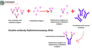
1. Double Antibody RIA
In this type of RIA second Antibody is added in the precipitation of main Ab and Ag and forms anti-immunoglobulin.
2. Coated-tube RIA
In coated tube RIA, primary Ab is coated in the tube. unbound Ag or Ab are present in the supernatant of the Solution which is removed.
Radioactivity is measured by a gamma counter in each type of RIA. Ag and Ab are specific for each other hence this test is very specific for diagnosis.
Methods
RIA is performed by stepwise method. These are the following steps of Radioimmunoassay
- Take a microtiter plate and it’s well fixed with the known concentration of antibodies.
- The radio-labeled antigen (hot antigen) was added to the well in excess quantity.
- The hot antigen will bind with antibodies. When the concentration of hot antigen is high, all the binding sites of antibodies will be saturated by the antigens. No antibody will be free. This time radioactivity is higher.
- Some radio labels are not bound with Ab. This unbound antigen is removed by washing through a buffer solution.
- Add the patient sample that contains cold or unlabeled antigens.
- unlabeled antigen competes with hot antigen. They replace hot Ag and bind with antibodies.
- This replacement of cold Ag releases Hot Ag away from the Ab. Then again the well to remove unbound Ag.
- A number of radiolabeled Ag binds to the Ab are decreased due to the addition of unlabeled antigens hence radioactivity is decreased.
- The radioactivity of the solution is measured by a gamma counter.
- The intensity of radioactivity is proportional to the concentration of Ag present in the patient sample.
- If the patient sample contains specific Ag for antibody. It will bind with Ab and form an Ag-Ab complex by replacing labeled Ag.
- Labeled Ag concentration is higher in the solution hence the radioactivity of the solution will decrease and the result will be positive.
- When radiolabeled Ag binds with Ab this time radioactivity of the solution is maximum. If the patient sample does not contain specific Ag for Ab. So they can not bind with Ab.
They cannot be replaced with labeled Ag. At this time radioactivity of the solution remains the same and the result will be negative.
Radioimmunoassay test
Through RIA we can perform both qualitative and quantitative tests. In qualitative tests, we can find only positive and negative results, and in quantitative tests, we can find a concentration of antigen.
RIA methods determine the concentration of nearly all of the hormones as well as many drugs, proteins, and other compounds.
A variety of compounds of serum or other biological fluids present in very small amounts can be estimated, e.g. Tumor markers like prostate-specific antigen (PSA), Alpha-fetoprotein (AFP), etc. Vitamins, drugs.
Immunoradiometric assay and radioallergosorbent test (RAST) are examples of RIA. In RAST we can determine the allergen which is causing the allergy.
Double antibody RIA
Q&A
1. Which radioisotope emits alpha particles?
Plutonium, americium, curium and californium radioisotopes emit alpha particles.
2. What is a radioimmunoassay?
Radioimmunoassay is a technique that determines the concentration of foreign Ag which causes diseases in our body. This technique is based on radioactivity.
3. Where does radioactive iodine come from?
Radioactive iodine comes as a byproduct of fission and also occurs naturally. Fission is a process that is involved in ground nuclear testing, nuclear weapon testing, and nuclear reactor operations.
4. Where is ionizing radiation found?
Ionizing radiation is found in medical instruments like CT scans, X-rays,s and some daily life gadgets like smoke detectors, etc.
5. How much radiation is an MRI?
MRI worked with the use of magnets and radio waves to create the images. It does not use radiation, X-rays and CT scans both use ionizing radiation but in MRI we are not exposed to radiation.