Introduction
Microscopy helps us to understand or gives access to see the direct images of various tissues, organisms, organelles, microorganisms, molecular assemblies, and proteins. It is an important, complementary technique to visualize the macro and/or microscopic structure and to assign structure to function and vice versa. The discovery of an electron microscope was not sudden, in the beginning, Roman people used lenses to see distant objects or to see large objects.
The oldest known lens in the history of science is the lanyard lens (discovered by researcher lanyard from 721 to 705 BC). The modern lens used for scientific research and experimentation developed in Florence (Italy), between 1280 and 1285. Simple microscopes are made from this formula.
Then various experiments were started to make advanced microscopes. Around 1590, Zacharias and his father Hans Janssen first invented the complex microscope using a pair of convex lenses. That is why Zacharias is called the father of microscopes. Many scientists then experimented with microscopes to create more advanced and complex microscopes that played a major role in the study of microbiology and science. Nowadays, depending on the structure of the instrument, there are two types of microscope, such as light microscope and electron microscope (1) & (4).
Electron microscope
An electron microscope uses electrons (negatively charged atoms) instead of light to magnify an object. The wavelength of the electron is much shorter thus can make very tiny things visible.
In 1931, Ernst Ruska, a German physicist, was the first to invent the electron microscope. At first, fine objects less than 0.2 micrometers could not be seen with the help of an optical microscope. After the invention of this microscope, particles less than 0.2 micrometers became visible.
An electron microscope can also experiment with an object 1 million times smaller than a human hair. Hence it is one of the most important discoveries in microbiology and scientific experimentation (2) & (3).
Creation of a magnified image with the help of electron microscope
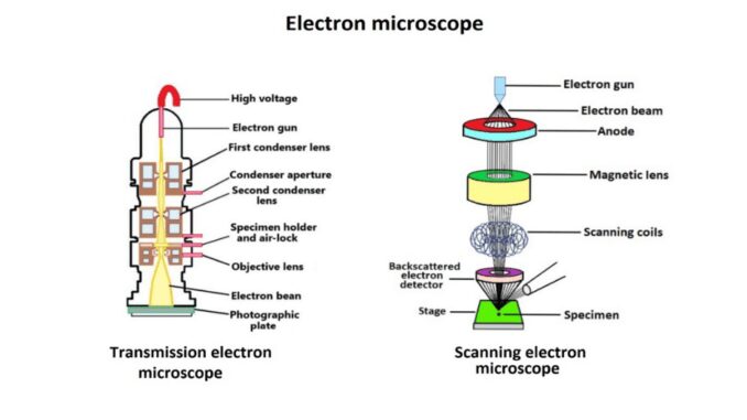
- Electron microscopes use high-voltage electrons instead of light. This high-voltage electron is formed by heated tungsten or field emission filaments which are electron sources.
- These electrons get accelerated in a vacuum and thus gain a positive electrical potential. The beam thus further becomes more focused by using a metal aperture and a magnetic lens.
- Hence forming a monochromatic beam and amplifying the image of the sample by using a magnetic lense.
- These microscopes use electromagnets instead of glass lenses as magnetic fields. The focus of the electron is also controlled by the electron lens. The power of this lens is reduced by increasing or decreasing the amount of electric current.
- The image obtained in this method is taken in a container part and then displayed. In this way, this microscope creates a magnified image (5).
Type of electron microscope
There are two types of electron microscopes according to their structural features.
1. Transmission electron microscope (TEM)
- In this device, electrons are emitted directly on the object in the form of rays by this an enlarged picture of the object is taken.
- The TEM is like a slide projector. In this microscope, some of the electrons are absorbed by the object or move to the other side. But the rest of the electrons that accurately create the image, in this case, this image is accepted.
- Much of this image is automatically trimmed before the required part is displayed. As a result, this device displays only a small portion of the enlarged portion. But this device presents the image in more detail.
- This type of device is considered high quality. The first electron microscope developed was a TEM (6).
2. Scanning electron microscope (SEM)
- This device uses a photographic plate on the back of the object.
- The resolution of SEM can be up to 1 nanometer. In some cases, the resolution may be lower than 1 nanometer.
- In this device, the photographic plate contains the magnetic pattern created by the electron particles hitting the object. As a result, a larger image of the whole object is obtained.
- Thus the image is obtained by SEM. This device produces 10,000,000 magnified images.
- SEM also forms three-dimensional images.
- In this microscope, thermal electrons are usually emitted by heating the tungsten filament. The thermal energy generated in this way is higher energy.
- The generated thermal electrons can be emitted with the help of a condenser lens in a diameter of 0.4 to 5 nanometers.
- These electrons gradually lose energy by acting on the object. The different types of symbols generated by this interaction are detected with the help of SEM detectors (2) & (6).
Parts of electron microscope
This device can be used to observe fine to very fine objects. These are electron guns, electron columns, electromagnetic lenses, and photographic plates. Other essential parts are high-voltage transformers, vacuum pumps, a water cooling system, a circulating pump, a refrigeration plant, and finally a filter system.
-
Electron gun
It is located at the top of the electron microscope. And is the main source of electrons used instead of light.
-
Electron column
It is an empty metal tube-type of a microscope. Electrons are spread by light elements like air. This part of the electron microscope is vacuumed.
-
Electromagnetic lens
A magnetic field is created under an electron microscope to determine the direction of motion of an electron particle. This magnetic field is called an electron lens. Electromagnets are used instead of glass lenses as the magnetic field of an electron microscope.
-
Photographic plate
The plate or screen behind an object observed by an electron microscope is called a photographic plate. This screen or plate contains the magnetic pattern created by the impact of the electron particle on the object. After projecting into a photographic plate, the object is transformed into an enlarged visible image.
-
High voltage transformer
Its function is to transform high-voltage currents for the electron gun and electron column.
-
Vacuum pump
The vacuum pump can maintain a high vacuum inside the electron column.
There are also some parts of an electron microscope such as the water cooling system, a circulating pump, a refrigeration plant, and a filter system. These parts also help in observing the enlarged image formed by an electron microscope (2) & (6).
Uses of electron microscope
Since the invention of the electron microscope, its use has been increasing day by day. Electron microscopes are important for showing the finest part of an object. The use of this microscope is very important in various scientific experiments.
- Due to the high resolution of the electron microscope (TEM), it is possible to observe the tiniest features of a sample.
- These microscopes are used to get detailed information about different types of organic and inorganic samples.
- Detailed analysis of different types of cell structures, small virus structures, biopsy specimens, etc can be done with this microscope.
- Electron microscopes (SEM) analyze in detail 3D images, topographical images, and data from various detectors.
- This device helps in the fast analysis of data due to the presence of strong and fast electron particles.
- Electron microscopes are used in physics, chemistry, microbiology, and various scientific laboratories.
- This instrument is widely used in medical and forensic labs.
- An electron microscope is used to examine microscopic particles.
- This microscope is used in gemology, metallurgy, nanotechnology, etc.
- Electron microscopes are widely used for the development of biochemical sciences, technologies, and industries.
- The crystal structure and the internal fracture of an object can be examined by an electron microscope (2) & (6).
The disadvantage of an electron microscope
Although the role of the electron microscope is significant in the history of science, there are some disadvantages in using this device.
- One of the disadvantages of an electron microscope is that it displays a small portion of the magnified part.
- This device is very large in size and therefore a specific large room is needed to store this device.
- Electron microscope requires a lot of money for proper storage and maintenance.
- This device cannot be used for very high-level research.
- More training is required to operate this microscope properly and to collect accurate samples from the microscope.
- Only samples of solid objects can be collected by electron microscope (SEM)
- As the electrons are scattered around in the air, the electron column of this microscope is vacuumed. For this reason, no living samples can be collected by an electron microscope.
- The specimen of the object collected from this device is white and black in color. So the images obtained from here have to be repainted later (5) & (6).
