

Introduction
A microscope magnifies and reveals fine details. This article is about different Microscope parts with labelled name and their functions, elucidating the instrument’s role in the scientific exploration and observation of minuscule structures.
Tasks that a microscope performs
- It should be able to produce a magnified image of the specimen.
- Better resolution
- Better contrast for a clear visibility.
Basic types of microscopes
Optical microscopes
Optical microscopes were highly popular during the nineteenth century. The best optical microscopes can resolve about 2000 Angstroms. Another name for optical microscopy is Light Microscopy.
They are used primarily by scientists to examine the microstructure of materials. The basic principles of microscopes involve image formation, magnification, and resolution.
Electron microscopes
Electron microscopes generate images with higher magnification and resolution than the light microscopes. The high resolution of electron microscopes results due to the short wavelengths of electrons. Such high resolutions enable electron microscopy to generate ultrafine images. There are two main types of electron microscopes:
Transmission Electron Microscopy
In transmission electron microscopy, the visible light ray is replaced by an electron ray and glass lenses for visible light. In the TEM system, an electron gun generates a high-energy electron beam for illumination.
Scanning Electron Microscopy
The scanning electron microscope is the most widely used type and generally examines structures by scanning the surface of materials. An SEM image is formed due to an electron beam that is responsible for scanning the surface area of the specimen. It thus creates a three-dimensional appearance of the image formed.
Stereo microscopy
It is a type of optical microscopy that uses two separate lenses to provide a three-dimensional view of the image. Stereo microscopes are used to view objects at different magnifications without changing lenses or adjusting the focus settings.
Applications
- The optical microscope allows researchers to observe living organisms at high magnification and study their structures.
- Optical microscopes are used by forensic departments to analyze evidence collected from crime scenes like hair strands, and fingerprints.
- An electron microscope plays a crucial role in studying nanoparticles and examining their behavior at an atomic level.
- Electron microscopy techniques are used for quality control processes in pharmaceutical industries.
- In semiconductor manufacturing processes, TEM is used highly for quality control purposes like detection of impurities or analyzing crystal structure.
- Scientists use TEM to study environmental samples such as air pollutants or soil contaminants present.
Common components of the microscope
Microscope parts labelled
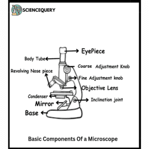
Main parts
Eyepiece
- The eyepiece is an important component of a microscope. It is generally used to view the primary image formed by the objective lens. The focusing of the eyepiece is a necessary step to observe images under a microscope.
- A good-quality eyepiece helps to provide clear images with minimal distortion and optimal brightness levels. A typical eyepiece has 10x or 16x resolution power whereas higher-powered ones consist of 20x – 45x powers.
Objective lens
- The objective lens of a microscope plays an important role in determining the quality and resolution of an image. The main aim is to collect light from an object and focus it onto the eyepiece.
- The numerical aperture of an objective lens varies from 0.16 to 1.40 depending upon the type of lens. A lens with higher magnification has a higher numerical aperture.
- There are two types of objective lenses: low power and high power. Low-power objectives have shorter focal lengths and magnify up to 20X. High-power objectives have longer focal lengths and have magnifying capacities of up to 1000x magnification.
Stage
The stage of a microscope helps to hold the specimen in place and allows for precise manipulation. Its main role is to hold the specimen in place to focus precisely.
Condenser
The condenser of a microscope is an important component that focuses light onto the specimen. It consists of several lenses and other components, such as diaphragms and filters. They control the amount of light passing through the objective lens. The condenser also helps to reduce aberrations in the image.
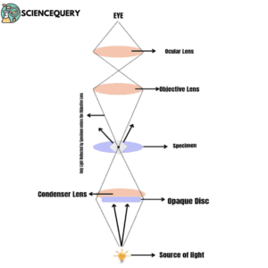
Illuminator
The illuminator or light source of a microscope is an important component that helps to provide the light source for viewing specimens. Different types of light sources used in microscopes depending on their applinclude halogen bulbs, LED lights, or fluorescent tubes.
Diaphragm
The diaphragm in microscopes is an important tool for improving image quality and clarity. With the help of a diaphragm, one can control the amount of light falling onto the specimen. It allows better resolution and contrast while viewing different samples. It controls the entire color contrast, allowing you to view the specimen in detail.
Coarse and fine focus knobs
The use of coarse and fine focus knobs in microscopes is an essential part. These knobs allow for precise adjustments to be made to the focus of the microscope, allowing users to view objects at different levels of magnification.
Coarse focus knobs are larger and control the movement of the stage up and down. It changes the distance between the objective lens and the specimen.
Fine focus knobs are smaller and usually found in front or behind one of the coarse focus knobs. They provide more delicate movements that allow for finer adjustments. Both knobs work together to achieve clear images by adjusting both horizontal (x-axis) and vertical (y-axis) positions simultaneously.
Base
It serves as the foundation upon which all the other parts are attached, including the stage, arm, and head. A heavy base will provide better stability to the entire instrument
Arm
The arm of a microscope connects the base and the head of the instrument. Metal arms provide more durability and stability, while plastic arms offer flexibility. Usually, microscopic arms are curved or angled to allow for comfortable hand placement during use.
Nosepiece
The nosepiece allows smooth rotation between objectives and maintains a safe distance from the stage.
Aperture
The type of aperture is determined by the type of objective lenses used. Different apertures use different objectives. Controlling these factors is crucial while using a microscope, as it directly impacts the image quality and accuracy.
Maintenance and Care Of Microscopes
Regular Cleaning
Microscopes need to be cleaned regularly with a soft muslin cloth. It requires proper disinfection as it comes in contact with live specimens that may contaminate the instrument.
Lubrication of components
Some parts of the microscope such as focus knobs, and diaphragms need to be lubricated periodically for easy movements.
Handling With Care
Microscopes are delicate and important tools in every pharmaceutical lab and molecular biology lab. Hence they need to be handled with care. It is recommended to handle the microscope only from its base or its arm.
Storage
The microscope should be stored in a clean, dry place to avoid any dust contamination. They should not be exposed to direct sunlight.
Calibration
Microscopes need to be calibrated periodically to achieve the precise results. The magnification settings must be calibrated to ensure accurate results.
Q&A
What are the 12 parts of a microscope?
The 12 main parts of a microscope include an eyepiece, objective lens, nosepiece, stage, condenser lens, apertures, illuminator, arm, base, fine focus knobs, coarse focus knobs, and diaphragm.
What are the 13 parts of a microscope and their functions?
The 13 main parts of a microscope and their functions are
Eyepiece: Also known as the ocular lens, which is used to view the specimen.
Objective lenses: Located on the revolving nosepiece and have different levels of magnification (4x, 10x, 40x, or 100x).
Revolving nosepiece: It helps in rotating the objective lenses for easy switching between magnifications.
Stage: A flat platform where slides containing specimens are placed for viewing.
Stage clips: Used to hold the slide in place on the stage.
Diaphragm: Located under the stage, it controls how much light passes through onto the specimen.
Condenser lens: Focuses light from below onto the specimen for better visibility.
Light source: Provides illumination for viewing specimens.
Courses adjustment knob: Moves either coarse focus or fine focus depending upon which one is being used at any given time.
Fine adjustment knob: Fine tunes focusing when using high power objectives.
Arm: Supports upper part of microscope above base, also serves as a handle while carrying scope.
Base: Bottom portion of microscope that provides stability and support.
Iris diaphragm lever: Controls the size of the opening in the condenser allowing more or less light into the system.
Mention the 13 parts of a compound microscope.
The 13 parts of a compound microscope include an eyepiece, body tube, coarse adjustment knobs, fine adjustment knobs, revolving nosepiece, handles, objective lens, condenser, inclination joint, mirror, base, stage, and substages.
What are the most important parts of a microscope?
The most important parts of a microscope include optical and non-optical parts. Optical parts of the microscope include the diaphragm, condenser, objective lenses, and ocular lenses. Nonoptical parts include the base, pillar, inclination joint, stages, and adjustment screws.
Summary
- There are different microscopic parts of a microscope.
- A microscope is a tool designed to magnify images and make fine details visible.
- Optical microscopes were highly popular during the nineteenth century. The best optical microscopes can resolve about 2000 Angstroms.
- The optical principles of microscopes include image formation, magnification, and resolution.
- There are two types of objective lenses: low power and high power. Low-power objectives have shorter focal lengths and magnify up to 20X. High-power objectives have longer focal lengths.
- Electron microscopes generate images with higher magnification and resolution than light microscopes.
- An electron microscope plays a crucial role in studying nanoparticles and examining their behavior at an atomic level.
- TEM is used highly for quality control purposes like detection of impurities or analyzing crystal structure.
- The stage of a microscope helps to hold the specimen in place and allows for precise manipulation.
- Diaphragms in microscopes are an important tool for improving image quality and clarity.
- The illuminator or light source of a microscope is an important component that helps to provide the light source for viewing specimens.
- Different types of light sources used in microscopes depending on their applications include halogen bulbs, LED lights, or fluorescent tubes.
- The nosepiece allows smooth rotation between objectives and maintains a safe distance from the stage.
- Microscopes are delicate and important tools in every pharmaceutical lab and molecular biology lab. Hence they need to be handled with care.
References
- https://books.google.com/books?hl=en&lr=&id=5YNTEAAAQBAJ&oi=fnd&pg=PP1&dq=microscope+parts&ots=Vgi9wq4l9H&sig=Aq4R6F8DI3gycdMv10J_UqJP6j4
- https://www.jstor.org/stable/44440748
- https://books.google.com/books?hl=en&lr=&id=zLxzEAAAQBAJ&oi=fnd&pg=PT140&dq=forensic+applications+compound+microscope&ots=7gpos5TVW7&sig=zfU3jbKMfC1rMYUPWIPeQX8-0yo
- https://link.springer.com/chapter/10.1007/978-3-031-08834-6_3
- https://link.springer.com/chapter/10.1007/978-3-030-64686-8_25
- https://www.academia.edu/download/64506885/Rodaks%20Hematology%205th%20Ed_booksmedicos.org.pdf#page=51
Written By: Sushmita Mukhopadhyay