

Introduction
What is dark field microscopy?
Dark field microscopy is a light microscopic technique, used to observe living cells and microorganisms without staining methods. This method involves illumination of samples from the side, rather than the front view. This technique is generally used to carefully study microscopic organisms like bacteria and protozoans.
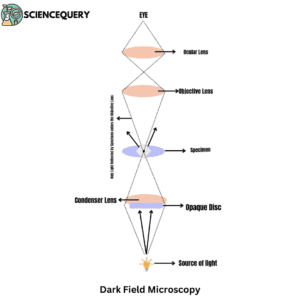
A special condenser is used that reflects light onto the specimen like a hollow cone at a particular angle. Dark field microscopy uses reflected light as its source of illumination and doesn’t allow light to fall directly on the objective lens. The specimen sample when placed on the stage, appears bright against the dark background.
Basic principle of dark field microscopy
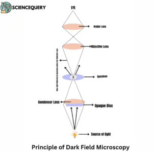
The technique generally uses a strongly focused light source that illuminates at certain parts of an object, creating a dark background. This allows small structures and organisms to be seen more clearly, making it useful for studying bacteria and other microscopic objects.
Difference between dark field and bright field microscopy
Characteristics |
Dark Field Microscopy |
Bright Field Microscopy |
Mode Of Illumination |
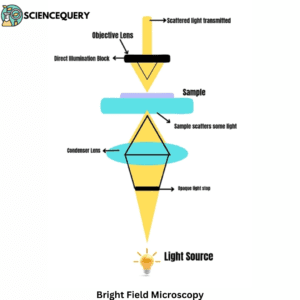
Dark field microscopy is an illumination technique used in the enhancement of unstained specimens. | Bright-field microscopy enables us to observe high-contrast images and to point out the fine details in specimens. |
Condenser Type |
A special condenser is used that reflects light onto the specimen like a hollow cone at a particular angle. | The condenser has adjustable elements like diaphragms that help in tuning the amount of light. |
Wave Source |
The light from the plane wave source is focused through an object. | An opaque disc is placed between the source and the condenser, blocking the middle of the beam. |
Diagrammatic illustration of both microscopy
Bright Field Microscopy Dark Field Microscopy
History or key features of the discoveries in dark field microscopy
- During the 1840s, the specimens used for light microscopy were diatoms. Diatoms belong to an algal species that possess intricate cell walls thereby making them ideal for research.
- The technique was first introduced by German physicist Ernst Abbe in the year 1873.
- This technique has been used for a variety of applications including medical diagnostics, material science research, and forensic analysis.
- Dark field microscopy is based on the principle that light can be scattered from particles or objects when illuminated at an oblique angle. This allows researchers to observe specimens without having to use stains or dyes.
- Hubert von Gruning then discovered staining techniques that made thin tissues easily viewable under a microscope even without specialized dyes.
- This technique has since found many applications in medicine like diagnosis of malarial infections and blood parasites.
- It helped in research into quantifying cell functionality, thus making it one of the primary tools utilized today. It is widely used for viewing living cells non-invasively in their natural environment.
Components
The components of a dark-field microscope include the following
1. Source of Illumination
This component uses angled illumination rather than regular transmission. It thus creates a ‘dark-field’ environment where everything outside focus becomes hazy due to the blurring.
2. Condenser Lens
The condenser lens moves up and down, allowing for alteration of the angles for optimal resolution.
3. Objective Lenses
These objective lenses magnify images by collecting the reflected beams from above or below the surrounding structures illuminated within the focus.
4. Eyepiece
The eyepiece is situated just below the barrier filter. It thus helps in minimizing the number of scattered rays entering the eye piece and provides better image resolution.
5. Dark field stop
The dark field stop is also called an annulus. It is generally a circular metal disk located at the base of the microscope that controls the amount of light entering through its central aperture.
How dark field works
- The dark field stop is responsible for blocking out bright light and allowing only low-intensity illumination to pass through it.
- It therefore results in a darker image with higher contrast.
- The darkness achieved by using a dark field stop allows greater visibility into deeply stained specimens.
- Proper adjustments are essential for obtaining maximum resolution from your specimen.
- It helps make objects appear lighter against a darker background, increasing visibility and higher detailing.
Sample preparation
- Cleaning the sample to wipe off any dust that may interfere with clear imaging.
- The sample is mounted on a slide using glycerol or oil immersion media.
- A thin layer of carbon film is put over the surface of the sample to reduce light scattering.
- Adjusting the illumination so that only scattered light is visible. It can be done by adjusting the condenser lens.
- Observing the specimen at different magnifications.
- Multiple exposures at different magnifications ensure to capture of details.
Advantages of Dark Field Microscope
- It allows for high-resolution imaging of small objects such as bacteria and viruses.
- Dark field microscopy also gives information about the size, shape, and orientation of particles.
Limitation of Dark Field Microscope
- It is used for opaque samples. It means that light cannot pass through them.
- Dark microscopy is good only for small, microscopic specimen studies rather than the larger particles.
Applications of Dark Field Microscope
- It helps in the clinical diagnosis of diseases like cancer proliferation, thus allowing a clear view of live cells.
- It helps in clearer analysis of biomolecules at nanoscale levels, widely used for genetic studies.
- Dark field microscopy is used to study the interaction of bioactive molecules to understand drug efficacy.
- This technique helps to find out the type of microorganisms suspended in liquids.
Summary
- Dark field microscopy is a light microscopic technique, used to observe living cells and microorganisms without staining methods.
- This method involves illumination of samples from the side, rather than the front view.
- It is generally used to carefully study microscopic organisms like bacteria and protozoans.
- The technique was first introduced by German physicist Ernst Abbe in the year 1873.
- Dark field microscopy is based on the principle that light can be scattered from particles or objects when illuminated at an oblique angle.
- Hubert von Gruning discovered staining techniques that made thin tissues easily viewable under a microscope even without specialized dyes.
- It helped in research into quantifying cell functionality, thus making it one of the primary tools utilized today.
- The dark field stop is responsible for blocking out bright light and allowing only low-intensity illumination to pass through it.
- Dark field microscopy is used to study the interaction of bioactive molecules to understand drug efficacy.
- The condenser lens moves up and down, allowing for alteration of the angles for optimal resolution.
- This technique helps to find out the type of microorganisms suspended in liquids.
- Dark microscopy is good only for small, microscopic specimen studies rather than the larger particles.
- The dark field stops, also called an annulus. It is generally a circular metal disk located at the base of the microscope that controls the amount of light entering through its central aperture.
- Dark field microscopy also gives information about the size, shape, and orientation of particles.
References
- https://pubs.acs.org/doi/full/10.1021/acs.analchem.0c04390
- https://www.sciencedirect.com/science/article/pii/S0927776514002860
- https://pubs.acs.org/doi/full/10.1021/acs.analchem.0c04390
- https://pubs.acs.org/doi/abs/10.1021/acs.analchem.6b02429
- https://www.sciencedirect.com/science/article/pii/S0255085721001997
- https://books.google.com/books?hl=en&lr=&id=caTUCgAAQBAJ&oi=fnd&pg=PR9&dq=dark+field+microscope+history&ots=_qT2rZBROF&sig=TwBHzXv1zFD8_syeffGCk6L3O-k
Written By: Sushmita Mukhopadhyay