

Know in one minute about Cornea vs Sclera
|
Introduction
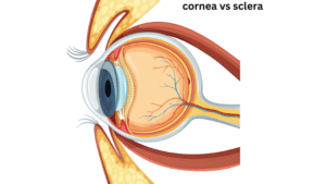
The cornea & sclera however these two parts of the eye stay adjacently but they are not the same. We can compare them in terms of their structure & functions. Thus, this article is about the cornea vs sclera.
The cornea is the clear translucent dome-shaped part present at the front of the eyeball. It covers the lens & the anterior chamber whereas the sclera is the continuous part that covers the whole eyeball. We call it the white part of the eye. It connects smoothly with the sclera and acts as a magnifying glass. Here we will try to be familiar with all the functions, roles, structures & differences of these two very important anatomical structures which give us the opportunity to read this article. Both of them are considered as the outer coat of the eye which essentially protects the structures inside it. They act as structural barriers against any outside danger & do perform many different important functions. Sclera maintains rotation of the eyeball, lubrication & all the optic nerve connections etc where the cornea functions in refractions of the light through the lens below it, etc.
Structure and Location of Cornea
Cornea is a transparent anterior layer of tissues which covers the frontal part of the eyeball covering the iris, pupil & lens. This is the area where we usually put our contact lenses.
It separates from the sclera at the corneoscleral junction. The cornea is composed of many unmyelinated nerve endings. That is those nerve endings that are not covered with myelin sheath. That’s why it is sensitive to touch, temperature, and chemicals. As transparency is of prime importance, the healthy cornea does not have or need blood vessels within it. A simple touch or a try will cause the closing of the eyelid involuntarily.
Layers of the human cornea
The human cornea is composed of five layers, from anterior to posterior the layers are
- Corneal Epithelium.
- Bowman’s Layer.
- Stroma
- Descemet’s layer
- Corneal endothelium.
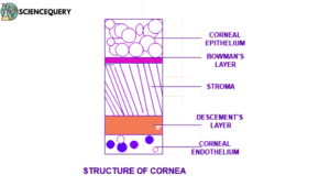
1. Corneal epithelium
The corneal epithelium is made up of epithelial tissue. The function of it is to form a barrier to protect the cornea, resisting the free flow of fluids from the tears.
2. Bowman’s Layer
This is the non-regenerating and most interestingly this is an acellular layer present between epithelium & stroma. Made up of collagen fibers & have a thickness of 8-10 μm.
3. Stroma
The thickest layer of the cornea is perfectly transparent. Posteriorly it is attached to descemet’s layer. It is about 200 μm thick, made up of layers of collagen fiber. This lamella are produced by keratocytes cells. The arrangement of each collagen fibrils is responsible for light scattering to the retina.
4. Descemet’s Layer
This is an extension of stroma which is made up of different types of collagen fibre. Posteriorly it is attached to the endothelium.
5. Corneal endothelium
This is the last layer of the human cornea, made up of endothelial cells & it is attached with aqueous humor (the fluid part present between the cornea & pupil). It mainly helps in solute transportation.
Functions of cornea
Cornea’s distinctive structure helps in the function & maintenance of the eye. Not only in terms of its ability to capture & process visual information also for the health of the eye.
Structure and Location of Sclera
Sclera is the tough, hardened fibrous & opaque outer layer that covers most of the eyeball. This is the whitish part that we can see from the outside along with the lens. It becomes thickened at the point surrounding the optic nerve at the back of the eye.
However it is referred to as white of the eye, and it has some pigmentation. Usually in children, it appears with a slightly blue hue as the structure is thinner. In adults due to fat deposition, it shows slightly yellowish in color. It also has blood vessels present at the surface of it.
Layers in the human Sclera
The sclera in humans has four distinctive layers.
Primarily sclera is made up of collagen fibers which give it elasticity & rigidity.
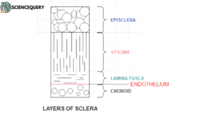
From outside to inside the layers of the sclera are arranged like this,-
1. Episclera
The outermost layer is made up of thin & loose connective tissue overlying it, also forming the sclera & conjunctiva. Later one performs the lubrication of the eyeball by producing mucus & tear. It’s basically a very thin mucous membrane.
2. Scleral Stroma
The medical term for it is substantia propria. Made up of collagen bundles, fibroblast, elastic fiber & ground substances. The ground substances are mainly glycoprotein & proteoglycans. Collagen bundles here are not arranged as regularly as in the cornea, that’s why the sclera is opaque.
3. Lamina fusca
The innermost layer of the sclera. Here melanocytes can be found abundantly.
4. Scleral endothelium
This is a simple squamous epithelium covering the gap present between lamina fusca & choroid, that gap is named suprachoroidal space.
5. Choroid
This is the vascular layer present between the sclera and retina. It generates the melanocytes to the lamina fusca & also helps in the nourishment of the outer retina.H.3.2.
Function of sclera
The primary function of the sclera is to protect the eye and maintain the shape of eyeballs.
Role of Cornea
- As the cornea has its unique dome-shaped convex structure it serves to focus & refract the light before it passes through the iris & lens to the retina.
- It also acts as a protective barrier to the internal structures of the eyeball.
- As it has dense nerve endings known as corneal nerves, it also triggers protective reflexes such as blinking, etc in exposure to any potential injury.
- Along with the lens, it also helps in the focus of the light, thus facilitating the vision.
In summary, the cornea’s functions of light refraction, protection, sensitivity, structural support, and contribution to the eye’s optical power are critical for maintaining vision and the health of the eye.
![]() Amazing facts about the human cornea
Amazing facts about the human cornea
It has a self-healing capacity. Shark corneas are used in human corneal transplantations.
Role of sclera
- The most crucial role played by the sclera is to provide protection to internal structures like choroid, retina, vitreous humor, etc.
- Its unique structure prevents the collapse of the eye from the socket and also works as an anchor for the extraocular muscles thus also helping in eye movement.
- Sclera has a very rich vascular network that provides nutrients & oxygen to the layers of the eye.
- The anterior part of the sclera has a slight depression known as “serial sulcus”, it helps to maintain eye pressure by draining the aqueous humor & it also accommodates the iris the shutter of our eye.
- It also helps minorly in light refraction, light enters through the cornea then onto the lens, and reaches the sclera before the retina. Due to this, the whitish color of it also refracts light a bit.
![]() Amazing facts about human sclera
Amazing facts about human sclera
We, humans, are the only beings with white sclera on earth. The Tenon’s capsule is a fascial sheet that forms the eye socket & it attaches to sclera anteriorly.
Comparison between cornea and sclera
Cornea |
Sclera |
| The transparent convex anterior portion of the outer fibrous coat of the eyeball. | The tough white fibrous outer envelope of tissue covers all of the eyeball except the cornea. |
| The cornea is the major refractor of the eye. | Sclera maintains the shape of the eyeball. |
| It covers the iris, lens & the pupil. | Covers all the internal parts of the eye. |
| ⅙ Part of the outer layer of the eye is covered by it. | ⅚ Part of the outer layer covered by the sclera. |
| Along with light refraction, it protects the eye by sensing any danger & using voluntary reflexes. | It helps to maintain the pressure inside the eye. |
| The epithelial layer receives all the nutrients from the tear and uses it. | Blood, oxygen, and nutrient supply are done by it. |
| Has many nerve endings which help to sense any danger & take defensive reflexes. | It helps to place the eyeball into the socket with the help of ocular muscles. |
| Has self-healing properties. | Don’t have any such property. |
- The cornea and sclera make up the outer tunic of the eye.
- Each of them is a connective tissue containing collagen fibrils embedded in a proteoglycan-rich extra-fibrillar matrix.
- Both tissues require strength to maintain the excess pressure within the eye and to resist external knocks and the forces applied by the extraocular muscles during eye movement.
Disorder or common condition affecting the cornea
Common corneal disorders are of various types. Small injuries like scratches and all can be healed by themselves over the course of time.
1. Allergies can occur which leads to conjunctivitis or red eye.
2. Corneal ulcers, a common disorder occur when an open sore develops in the cornea. Cause of it can be various like any mechanical injury, drug abuse, misuse of contact lenses, etc.
Usually treatable with antifungal eye drops and antibiotics.
3. Bullous keratopathy, a common disorder seen in elderly persons. The cause of it is edema in the cornea. A blister-like appearance is seen in the cornea composed of blurry liquid.
Treatable by topical hypertonic solutions.
4. Herpes zoster ophthalmicus is a reactivated infection of the eye caused by the varicella-zoster virus, the virus that causes chickenpox and shingles.
Keratitis
1. Interstitial keratitis, is a serious corneal disorder where the middle layer of the cornea becomes inflamed. Usually occurring in children, bacteria-caused syphilis is the major contributor. Blurry vision & sensitivity to light are the main symptoms.
Treatable with corticosteroid eye drops.
2. Superficial punctate keratitis is an eye disorder caused by the death of small groups of cells on the surface of the cornea.
Caused by viral or bacterial infection, even dry eyes can cause it.
Treatment depends on the cause
Autoimmune diseases
- Cogan Syndrome, It’s a rare autoimmune disease. An autoimmune disorder is a malfunction of the body’s immune system that causes the body to attack its own tissues.
- The usual symptoms are redness, dryness & loss of vision.
- Treatments involve corticosteroid eye drops, sometimes corticosteroids given by mouth but as it’s an autoimmune disease it’s difficult to cure completely.
Disorders or common conditions affecting sclera
We can divide scleral inflammation broadly into two categories
1. Scleritis
2. Episcleritis
Both of them are recurrent inflammation.
Scleritis
It is a severe inflammation of the tissues of the sclera. This condition is characterized by edema & cellular infiltration.
Direct damage to the ocular nerves associated with sclera cause severe pain in scleritis, also there are ocular muscles attached to the sclera, so eye movement can cause severe pain in this infection.
The actual cause of the disease is unclear but usually associated with rheumatoid arthritis, connective tissue diseases, “Sjögren’s syndrome”, medicines used to treat or prevent bone disease, trauma, etc.
Three types of scleritis are present
- Diffuse scleritis: Here the infection is scattered all across the sclera. It’s the most common type.
- Nodular scleritis: Nodular scleritis is concentrated in one spot of the sclera. You can usually see the lump (nodule).
- Necrotizing scleritis: This 9s the most severe form of scleritis. It can destroy eye tissue and even result in the loss of your entire eye.
Since sclera is dependent on episclera providing a response to the inflammatory stimulus, scleritis is always accompanied by overlying episcleritis.
Episcleritis
It refers to inflammation of the episclera, which is a clear layer on top of the white part of the eye.
Two types of this condition occur
- Simple – redness in a section or into the whole eye with minimal discomfort.
- Nodular- Bumps can be seen surrounded by dilated blood vessels, causing discomfort.
There’s a clear layer outside of the episclera called the conjunctiva.
Also known as Pink eye, a very common disorder. Caused by many reasons including viral, bacterial, allergic, or chemical injection
Q&A
1. Does the cornea cover the sclera?
The cornea does not cover the sclera. Because both of them cover the eyeball like the skin of a balloon covers the air inside it. Also, the cornea and sclera have distinct borders between them.
2. Cornea vs Sclera vs conjunctiva
| Cornea | Sclera | Conjunctiva |
| The cornea is the transparent outermost layer responsible for focusing light onto the retina. | Sclera is the tough outer layer providing structural support for the eye & known as the white of the eye. | Conjunctiva is a thin membrane that covers the inner surface of the eyelids and the front surface of the sclera, helping to protect and lubricate the eye. |
3. Does the cornea cover the entire eye?
No, the cornea is the transparent layer that covers the front part of the eye, covering the iris and lens. It covers roughly ⅙ part of the eye.
4. Is the cornea part of the sclera?
The cornea and sclera are distinct and separate structures in the human eye. Both of them are part of the external covering of the eye, but they are not the same. They are adjacent to each other and together form the outer covering of the eye, they are not physically or anatomically part of each other. The boundary between the cornea and sclera is known as the limbus.
5. What is the difference between cornea and sclera?
The main difference between these two is, the cornea is the frontal translucent cover of the eyeball which refracts the light to the retina. Where sclera is the white opaque part that covers the rest of the eyeball and mainly helps in nourishment & gives protection to the eyeball.