Introduction
Veins are a type of blood vessels in the body that carry blood. It is elastic and muscular in nature. Veins collect blood from various parts of the body and empty in the heart. All veins except the pulmonary vein carry deoxygenated blood. Veins have a low blood pressure system (5 to 10 mm of Hg).
Definition
A vein is a vessel that conveys blood away from an organ towards the heart. Veins are formed as a result of the union of venules. Venules are formed due to the union of capillaries.
Functions
1. Carry blood
Veins carry deoxygenated blood from the capillaries back to the heart. Through pulmonary circulation, this deoxygenated blood reaches the lungs. Thus carbon dioxide is got rid of the body.
2. Regulate pH
It regulates blood pH by transporting carbon dioxide.
3. Maintain circulation
Maintains continuity in circulation by returning the blood to the heart.
Blood reservoir
It acts as a blood reservoir or capacitance vessel. It accounts for about 70 % of the total blood volume at a time.
Significance
- The presence of valves ensures blood flow towards the heart only.
- The veins are near the skeletal muscles. As the muscles contract, the blood keeps moving.
- The respiratory pump helps the blood to go from the pulmonary and systemic circuit to the heart.
- Due to thin tunica media, it is innervated by the sympathetic nerves, causing venal constriction. Thus blood flow is ensured in the veins towards the heart.
Types
The following types of veins are found in the body
1. Deep veins
These are located within muscle tissues. They have a corresponding artery nearby.
2. Superficial vein
These veins are closer to the skin’s surface. They don’t have corresponding arteries.
3. Pulmonary veins
These veins arise from each lung (two sets ) and enter the left atrium of the heart. They bring oxygenated blood from the lungs to the heart. [This is an exception; as it carries oxygenated blood].
4. Systemic veins
They are located throughout the body. It is of three categories. They are: the heart veins, veins of upper body parts, and veins of lower body parts. It carries deoxygenated blood from all body parts back to the heart.
5. Portal vein
These veins start and end in capillaries. Such a circulation of blood is called the portal system. It is seen in the liver ( hepatic portal system ) and the hypophyseal portal system in the brain.
Structure
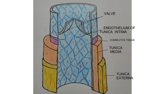
The average diameter of the veins ranges from 5 mm to 3 cm. It is composed of three layers
1. Tunica external
Outer thick layer of the vein wall. It is made of collagen tissue and is protective in function. Also non-elastic in nature. It contains tiny blood vessels called vasa vasorum. It supplies blood to the veins of the blood.
2. Tunica media
It is the thin and less muscular middle layer. Collagen is also present in this layer.
3. Tunica interna
This is the innermost layer; made of endothelium. It surrounds the lumen and minimizes the friction to the flowing blood. The endothelium folds inwards into flap-like structures called valves. These prevent the backflow of deoxygenated blood towards the organ from where they have been collected.
Diseases associated with veins
1. Deep vein thrombosis
A blood clot forms in a deep vein. This clot can travel to the lungs causing pulmonary embolism.
2. Varicose veins
Superficial veins near the surface of the skin swell. This is caused due to breakage or weakening of the valves. Thus blood collects or flows backwards.
3. Chronic venous insufficiency
This is caused when both deep and superficial veins’ valves malfunction. It’s similar to varicose veins but more symptoms like coarse skin texture and ulcers are seen.
4. Hemorrhoids
These are painful, swollen veins in the lower portion of the rectum and anus. It is caused by straining during bowel movements.
Difference between superior vena cava and inferior vena cava
Basis of difference |
Superior vena cava (SVC) |
Inferior vena cava (IVC) |
| 1. Location and passage to the right atrium | Located above the heart, posterior to the first costal cartilage. Then descends vertically to the trachea and aorta to enter the right atrium. | Originates at the lower back where common iliac (leg) veins join at the level of the fifth lumbar vertebrae. Runs under the abdominal cavity along the right side of the vertebral column to enter the right atrium. |
| 2. Functions | It returns deoxygenated blood from the head, neck, chest, and upper limbs to the heart. | Returns deoxygenated blood to the heart from all organs situated below the diaphragm. |
| 3. Structure | Valves are absent. Long vein with 27 to 36 mm diameter. | One valve (Eustachian) is present where IVC enters the right atrium. Short vein with 18 to 22 mm in diameter. |
| 4. Regulation of blood flow | Blood flow is regulated through the contraction of the muscular walls of SVC and gravitational force. | 4. Blood flow is regulated by the pressure from breathing and contraction of the diaphragm. Sympathetic nerve stimulation, and muscle contraction also help in blood flow through IVC. |
| 5. Veins draining | Veins draining into SVC are radial, ulnar, subclavian, brachiocephalic, and jugular veins. | Veins draining into IVC are lumbar, hepatic, suprarenal, and gonadal veins. |
| 6. Diseases | SVC syndrome is caused by lung cancer and lymphoma. | IVC syndrome is caused by tumors, deep vein thrombosis, and kidney diseases. |
Q&A
1. Where are the biggest veins in your body?
Superior and inferior vena cava .
2. What is the difference between arteries and veins?
Arteries are blood vessels carrying oxygen-rich blood away from the heart to the body. [except pulmonary artery]
Veins are blood vessels that carry deoxygenated blood from the body to the heart. [except pulmonary vein]
3. What is the main cause of varicose veins?
Due to improper working of valves of the vein.
4. What are the three layers of veins?
The three layers are tunica externa, tunica media, and tunica intima.
Summary
- Veins are blood vessels of the cardiovascular system. They carry deoxygenated blood from the cells, tissues, and organs to the heart. It constitutes a low blood pressure flow system. Veins are the blood reservoirs of the body.
- Veins arise from the union of venules. Venules are formed when capillaries converge. Veins have larger lumen than arteries. It is lined by endothelium forming the innermost layer tunica interna.Extensions of endothelium form valves to prevent backflow of blood. The tunica externa is the outermost thick layer made of collagen. In between occurs tunica media a thin layer of smooth muscle.
- Veins are of different types. The pulmonary veins bring oxygenated blood (except) from the lungs to the heart. The systemic veins occur all over the body; collecting deoxygenated blood and returning it to the heart.
- The superior vena cava collects deoxygenated blood from the upper parts of the body above the diaphragm. The inferior vena cava collects deoxygenated blood from all organs located below the diaphragm.
- The malfunctioning of the valves causes disorders like varicose veins, deep vein thrombosis, etc.
References
1. Human Physiology by C C Chatterjee (Vol I).
2. Human Physiology by C C Chatterjee (Vol II).
