Introduction
There are various species of animals in this world. Every animal has a number of organs that collect special stimuli from the environment and transmit them to the brain through specific nerves. All these organs are called sense organs. The five special sensory organs of the human body are the eye, ear, nose, tongue, and skin. Each organ has certain characteristics. For example, the ear helps to hear, the nose helps to smell, the tongue helps to taste, the skin helps to feel and the eyes help to see. Among these organs, the eye is the visual organ (1).
What is the eye?
The eye is a wonderful organ that allows us to see everything around us. When there is light in front of the eyes, the eyes respond and help us to see. The information that enters the human eye first reaches the brain through the optic nerve located in the eye. The brain then thinks about that information and helps people to make decisions. For example, if a bird flies towards a person, then the person moves away. In this case, the human eye sees the bird and sends a message to the brain through the nerves. And the brain is deciding the person to move on from there. About 95% of the world’s animals have eyes. The eyes of some animals can perform very simple functions, such as responding to light and darkness. But the eyes of some animals determine not only light and darkness but also color and depth.
Some animals have the same eye position as humans, with two eyes very close together so that the depth of vision is greater. But the eyes of some animals are quite far apart and are on opposite sides of the head. This makes it easier to find their prey. Such as rats.
Different members of the animal kingdom have different eye structures and functions. Such human eyes are different from the eyes of a fly. The fly’s eye can easily detect rapid movement, which the human eye cannot. The eye is the light-sensitive organ in the animals (3) & (4).
Definition of eye
The special organ by which animals receive luminous stimulus from the environment and see the outside world is called the eye. The human eye is the most valuable and sensitive organ. The eye is an important part of the human body, without which the human being is completely useless (3).
Structure of the human eye and its function
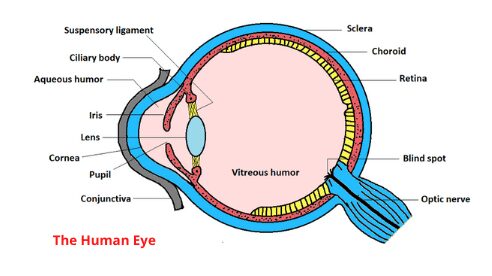
The eye is the human sense of sight. The human eye is located in front of the brain. Each eye has a specific structure. Each eye consists of an eyeball, a pair of eyelids, and a lacrimal gland. The main parts of the eyeball are conjunctiva, cornea, iris, lens, sclera, choroid, and retina. The different parts of the eye are described below (1).
1. Eyelids
The eyes are covered with a circulating upper eyelid and a lower eyelid. At the edge of the eyelids, there is an eyelash.
Function
The main function of the eyelids is to protect the eyes from external injuries and dust (2) & (4).
2. Conjunctiva
It is a kind of transparent thin covering located just outside the eyeball. It is located between the cornea and the eyelids (2) & (4).
Function
Its main function is to protect the inner part of the eye and the cornea (2) & (4).
3. Cornea
It is the transparent layer in front of the eyeball. The conjunctiva is on the cornea. The cornea is slightly thicker, whiter, clearer, and fibrous than the sclera. It is the anterior part of the eyeball associated with the sclera. It is located 1/6 of the space in front of the eye. An eye transplant is a corneal transplant (2) & (4).
Function
The cornea helps light to enter the eyeball. It acts as a refractory agent (2) & (4).
4. Iris
Iris is a black, opaque, round thin membrane with a hole in the middle. It is located behind the cornea and in front of the lens. Iris is the colorful part of the eye that looks a lot like a ring. It is of different colors, such as brown, green-blue, etc. (2) & (4).
Function
The main function of the iris is to increase the diameter of the pupil and control the entry of light into the eyeball (1).
5. Lens
It is the next bi-convex transparent part of the iris. There is no blood supply in the lens. With the help of the lens, people can easily see near and far objects. The lens can be compressed and stretched by the iris. It is made with crystalline proteins (2) & (4).
Function
The lens plays a major role in refraction. The lens also focuses light rays on the retina. (2) & (4).
6. Sclera
The sclera is the outer covering. It is a thick, white, opaque, and fibrous coating. The sclera is located at the back of the eyeball.
Function
The main function of the sclera is to protect the eye from external injuries and to give the shape of an eyeball (2) & (4).
7. Choroid
This is the next layer of the sclera. It is the middle layer located behind the eyeball. Because of melanin pigment, this part is black. It has an iris and lens inside. It is a layer of a dense pigment (3).
Function
The choroid protects the retina and prevents the reflection of scattered light. It supplies blood to the retina (3) & (4).
8. Retina
The retina is the next nerve layer in the choroid layer. It is the inner membrane at the back of the eyeball. This layer is made up of two types of nerve cells rod cells and cone cells. It is the light sensory part of the eye. It converts light rays into electrical signals and sends them to the brain through the visual nerves. Because of the presence of rod cells, people can recognize different colors and distinguish between them.
Function
The retina helps in the formation of reflections of objects. Rod cells and cone cells located in the retina act as light and color receptors. Rod cells are capable of absorbing soft light and color and cone cells are capable of absorbing bright light and color (2) & (4).
9. Pupil
The pupil is located at the back of the iris. Its size is controlled by the iris. This is the open part of the middle of the iris.
Function
Light enters the eye through the pupil (3).
10. Aqueous humor
It is a light aqueous mixture of sugars, proteins, and various salts. It is located between the lens and the cornea.
Function
Aqueous humor maintains the internal pressure of the eye and provides nutrition (2) & (4).
11. Vitreous humor
Vitreous humor is a jelly-like component of sugars, proteins, and various salts. It is located between the lens and the retina.
Function
It acts as a refracting agent and helps to maintain the internal pressure balance of the eye (2) & (4).
12. Lacrimal gland
A lacrimal gland is a number of tubular small almond-shaped glands. It is located almost in the middle on the inside of the upper eyelid. The fluid that comes out of the lacrimal gland is called tears. The secretion of the lacrimal gland is carried through the ducts and spreads over the conjunctiva and keeps the eyes wet (1) & (4).
Function
It helps to keep the eyes moist. When dust and sand fall on the surface of the eyes, tears help to clear it. Sodium carbonate and sodium chloride in the tears act as disinfectants (2) & (4).
13. Ciliary body and suspensory ligament
The ciliary body consists of ciliary muscles and a ciliary amplifier. And the suspensory ligaments are the fine fibers emitted from the ciliary muscle. These two parts of the eye are located between the choroid and the iris.
Function
The suspensory ligaments hold the lens. And the ciliary body helps in the accommodation (4).
A few more parts of the eye are the blind spot, yellow spot, and the optic nerve.
The blind spot is located on the retina on the opposite side of the pupil. There is no reflection formed at the blind spot.
The yellow spot is also located on the retina on the opposite side of the pupil. The reflection formation at the yellow spot is the best.
The optic nerve is the second cranial nerve in humans. Through this, the light sensation from the eyes reaches the brain (4).
Eye accommodation
The ability of the lens to control the focal length of an eye to see objects at any distance is called eye accommodation.
Accommodation method
1. As the ciliary body of the eye expands, the curvature of the lens decreases, and the lens becomes narrower. As a result, the focal length of the lens increases, and the reflection of the object is formed correctly on the retina. And people get to see the object.
2. When the ciliary body is compressed, the curvature of the lens increases, and the lens becomes thicker. The focal length of the lens decreases as the lens becomes thicker. As a result, the reflection of a nearby object falls precisely on the retina, and people can see the object.
Example
The accommodation allows the driver to see distant traffic signals by reducing the curvature of the lens of the eye while driving. Accommodation allows drivers to see any vehicle in front of the vehicle by increasing the curvature of the lens of the eye while driving (1) & (3).
Vision errors and correction methods
The following defects are seen when the accommodation capacity of our eyes is reduced.
1. Myopia
If the size of the eyeball is larger than normal, the reflection of the object is not formed in the retina. It is formed before (in the part of the vitreous humor). As a result, distant vision is disrupted. This abnormality of the eye is called myopia. This defect can be removed by using concave lens glasses (2) & (3).
2. Hyperopia
When the size of the eyeball is smaller than normal, the reflection of the object is formed behind the retina. As a result, the near vision is disrupted. This defect of the eye is called hyperopia. This defect can be removed by using convex lens glasses (3).
3. Presbyopia
The contraction- expansion of the lens of the eye of a 40-year-old man is reduced. This abnormality of the eyes is Presbyopia. In this case, distant objects can be seen, it is difficult to see near objects. This problem is removed by using convex lens glasses (2).
4. Cataract
With age, the lens or cornea gradually becomes opaque and vision becomes faint, called cataracts. Surgical replacement of the lens or cornea is the only cure (4).
