Introduction
The digestive system in frog is a complex network of organs and glands. They help in ingestion, digestion, absorption, assimilation, and egestion.
Frogs are carnivorous animals. They feed on earthworms, spiders, snails, and all types of insects. The prey is captured and swallowed whole. In this topic, we will know in detail about the digestive system of frogs.
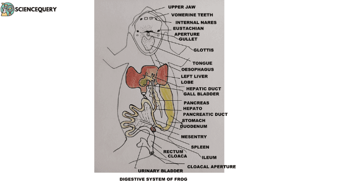
The digestive system of a frog comprises:
1. Alimentary canal
2. Digestive glands
Alimentary canal
It is a coiled tube, starting from the mouth and opening in the cloacal aperture.
From the histological point of view, this canal is made up of four layers. They are :
1. Serous coat – Outermost thin layer
2. Muscular layer – Comprises outer longitudinal muscles and inner circular muscles.
3. Submucous coat – Comprises connective tissue matrix with blood and lymphatic vessels.
4. Mucous coat – Innermost layer facing the lumen. It comprises epithelial cells and glands in certain regions.
The alimentary canal has the following parts:
- Mouth
- Buccal cavity
- Pharynx
- Oesophagus
- Stomach
- Small intestine
- Large intestine
- Cloaca
Mouth
- It is a wide aperture bound by the upper and lower lips. Lips are immovable.
- The mouth leads to the buccal cavity.
Buccal Cavity
- It has an upper jaw and a lower jaw.
- The upper jaw is immovably articulated with the skull; same as in higher vertebrates.
1. Teeth
- A row of small uniform hook-like pointed teeth are present in the upper jaw. These are maxillary teeth. It is used for holding onto the prey and not for chewing of food.
- Present on either side of the median line of the roof of the buccal cavity is the vomerine teeth. These teeth prevent the escape of prey.
- Teeth are homodont (similar in shape) and acrodont (attached to bones). During the lifetime of the frog, these teeth are replaced many times so they are also called polyphyodont.
- The tooth has two parts –the crown and the base. The base is attached to the jaw bone and is made up of a bone-like substance.
- The crown is made up of a shining white hard layer – enamel. Beneath the enamel is the dentine.
- The interior of each tooth is filled with a pulp cavity. It is highly vascular and contains odontoblasts cells that help in the growth of teeth.
2. Internal nostrils
- The roof of the upper jaw has openings of internal nares, connecting with the nasal cavities. The respiratory gases pass to and from the buccal cavity during respiration.
3. Eye bulges
- Impressions or bulges of the eyeball are seen behind the vomerine teeth. The eyes are retractable . After the prey is caught , the eyes close. This pushes the eyes downward and the prey is pushed towards the pharynx into the oeophagus.
4. Eustachian opening
- The opening of the Eustachian tube connects the pharynx with the middle ear. It is responsible for equalizing air pressure on both sides of the eardrum.
5. Tongue
- The floor of the buccal cavity has a fleshy, sticky, muscular tongue. It is attached in front to the inner border of the lower jaw and free behind.
- The tongue is bilobed at its free end. The upper surface of the tongue bears taste buds in the form of papillae and mucous glands. the secretion of the mucous glands keeps the tongue sticky. The tongue can be thrown out and retracted suddenly to capture and engulf prey. The protractor and the retractor hypoglossus muscle bring about this action.
- The tongue movement is also brought about by the sudden flow of lymph from the sublingual lymph sinus. It is situated beneath the fixed end of the tongue.
- The lower jaw is hinged and freely movable.
- Salivary glands are absent.
Pharynx
The buccal cavity leads to a narrow pharynx. It contains :
- Glottis – a slit-like vertical aperture that leads into the respiratory passage.
- Gullet – a transverse aperture leading into the oesophagus. It opens only at the time of swallowing the prey.
- In male frogs, vocal sacs are present on either side of the floor of the pharynx. The males use these sacs to attract female frogs during mating.
Oesophagus
A broad , tubular short part beyond the gullet . It is because of the absence of neck but highly distensible.
- The inner lining is thrown into numerous longitudinal folds . This allows for expansion of oesophagus during the passage of ingested food.
- There is no demarcation between the oesophagus and the stomach.
Stomach
A wide tube between the oesophagus and the intestine.
- It is located on the left side of the body cavity. It is attached by the dorsal mesentery to the dorsal body wall.
- The mucous coat of the stomach contains tubular gastric glands. It secretes digestive enzymes for the digestion of food.
- The anterior part of the stomach is expanded and is called the cardiac part.
- The posterior part is short and narrow, known as the pyloric part.
- The internal lining of the stomach has numerous longitudinal folds. This allows for the expansion of the stomach. These folds thin out and disappear at the cardiac part. It converges in the pyloric part.
- A constriction with a ring muscle is seen between the stomach and the intestine. This is the pyloric sphincter which regulates the passage of food from the stomach to the intestine.
Intestine
The stomach leads to the next longest tubular part of the alimentary canal; the intestine. It has two parts :
1. Small intestine
It lies in several loops held in position with the body wall with mesentry.
- The anterior short part ( U –shaped ) is called the duodenum. It receives duct from the liver and pancreas.
- The mucous coat of the ileum has numerous finger-like projections called the villi. Intestinal glands are also present which secretes intestinal juices.
- The ileum is extensively coiled. At its lower end, it leads to the large intestine.
2. Large intestine
- A short, broad tubular part which is also known as the colon is divided into rectum and cloaca.
- The cloaca receives openings from the ureters, urinary bladder, and oviducts in females.
- The cloaca opens out to the exterior by a vent or cloacal aperture.
- Through this aperture urine, gametes, and fecal matter are eliminated from time to time.
Digestive gland in frog
The liver and the pancreas are the important digestive glands, besides gastric and intestinal glands.
Liver
- This is the largest gland found in the body. It is reddish brown, located close to the heart and lungs.
- It consists of two lobes – right and left. A narrow bridge of liver tissue connects the two lobes.
- The left lobe is again subdivided into two lobes.
- Between the right and the left lobes and on the surface of the liver is found the gall bladder. It is a thin-walled, round, large greenish sac storing bile secreted by the liver cells.
- The bile secreted from the liver comes out from the hepatic duct and is stored in the gall bladder. It’s a greenish-colored secretion; alkaline in nature.
- The cystic duct from the gall bladder and the hepatic duct from the liver unite to form a common bile duct.
Pancreas
- This is the second largest digestive gland. Irregular in shape and lies between the stomach and duodenum. It consists of many small lobes.
- The common bile duct passes through the pancreas where it receives numerous pancreatic ducts. So it is also called the hepatopancreatic duct, which opens into the duodenum.
- The pancreas is both an exocrine and endocrine gland. The exocrine part secretes pancreatic juice.
- The endocrine part has Islets of Langerhans, which secrete the hormone insulin.
Physiology of Digestion
The food of toads consists primarily of insects. Tadpoles are herbivores feeding on aquatic plants. Vitamins, minerals, and water are absorbed as it is. Carbohydrates, proteins, and fats are complex organic compounds that are broken down by enzymes in digestive juices.
Ingestion
- Food is taken through the mouth. The sticky tongue captures the prey. It is put into the buccal cavity and the mouth is closed.
- The food is mixed thoroughly with the mucus in the buccal cavity.
- Through peristalsis (wave-like motion of the alimentary canal) the food moves into the oesophagus, then into the stomach.
Digestion
In Mouth
- Salivary glands are absent in the mouth of a frog. So no enzymatic digestion takes place here.
- The mucus of the buccal cavity and oesophagus moistens and lubricates the food.
In Stomach
- The parietal glands or oxyntic cells in the gastric mucosa secrete hydrochloric acid.
- The peptic cells secrete the enzyme pepsinogen ( inactive state).
- Pepsinogen is activated to pepsin( active) in the acidic medium. It acts on the protein of the food and converts into proteases and peptones.
- The carbohydrates and the fats are not digested here.
- After remaining for two to three hours in the stomach the half-digested food (chyme ) passes to the duodenum.
Intestinal digestion
Small Intestine
- In the duodenum the acidic chyme comes in contact with the bile, pancreatic juice, and intestinal juice.
- The acidified chyme stimulates the mucosa of the duodenum to secrete the hormone secretin and cholecystokinin.
Bile
- Choleocystokinin travels through the blood and reaches the gall bladder. It causes the gall bladder to contract and release bile through the hepatopancreatic duct.
- The alkaline bile neutralizes the acid of chyme. Bile has no digestive enzymes.
- It emulsifies fats, stimulates peristaltic action, and activates pancreatic lipase.
- Pancreatic Juice:
- Secretin stimulates the pancreas to secrete pancreatic juice. The pancreatic juice has three enzymes – trypsinogen( inactive state), amylase or diastatic enzyme, and lipase. They hydrolyze food in an alkaline medium.
- Trypsinogen is activated by enterokinase to trypsin. The trypsin then converts the peptones into soluble amino acids.
- Amylase acts on starch to give maltose.
- Lipase acts on fats to give fatty acids and glycerols.
Intestinal juice
Enterokinin
- Hormone activates the secretion of intestinal juice which is known as succus entericus.
- The succus entericus or the intestinal juice contains several enzymes like enterokinase, erepsin, maltase, lipase, and sucrase. These act on carbohydrates, proteins, and lipids.
- Erepsin converts polypeptides to amino acids.
- Maltase converts maltose to glucose.
- Lipase converts fats into soluble fatty acids and glycerol.
- Sucrase acts on sucrose to give simple sugar glucose.
Large Intestine
Here reabsorption of water and also the preparation and storage of faeces take place.
Absorption
- The final products of digestion are absorbed through the walls of the small intestine.
- The presence of villi-like processes increases the surface area of absorption.
- The epithelial lining absorbs water, mineral salts, and other nutrients through an osmotic gradient.
- Carbohydrates are absorbed as glucose and fructose. Proteins are absorbed as amino acids. They are taken up in the blood capillaries in the folds.
- The nutrients finally reach the liver through the hepatic portal vein.
- Fatty acids and glycerol pass into lymphatic capillaries ( lacteal) and then to veins.
Assimilation
- The absorbed food is used for the liberation of energy during respiration. It is also used as a part of the structure building of the animal body.
- Excess glucose is stored as glycogen in the liver or skeletal muscles or as fats in adipose tissues.
- Amino acids are utilized for growth and repair of tissues.
- In another route, amino acids can be deaminated to form urea and excreted with urine.
Egestion
The indigested food enters the rectum by peristalsis for storage and formation of feces. Old epithelial cells, leucocytes, bile pigments ,and bacteria are removed through feces periodically through cloacal opening.
References
Introduction to Biology of Animals – Volume -II by Sinha, Adhikari, Ganguly.
