Introduction
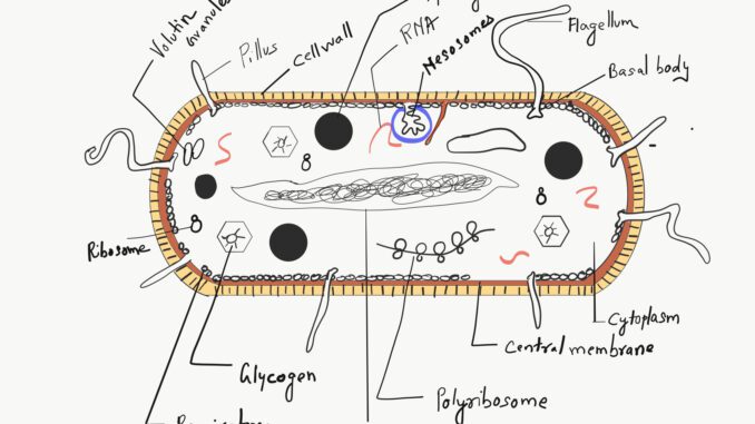
Microorganisms are categorized under bacteria, viruses, fungi, and mycoplasma. Some of the microorganisms are disease-causing some are saprophytes or some are heterotopic in nature. Bacteria are cosmopolitan, unicellular, rigid cell-walled, prokaryotic microorganisms. They are simple in structure cause diseases in plants and animals. The following article is about gram-positive and gram-negative bacteria and their features.
Characteristic features of bacteria
1. Bacteria are found almost everywhere, they are simple, unicellular, and primitive organisms.
2. The bacteria are autotropic, parasitic, saprophytic, and symbiotic in nature.
3. Bacteria are prokaryotic in nature and do not have a well-developed genetic system.
4. It lacks cellulose but has a rigid cell wall composed of mucopeptide.
5. Bacteria are both motile and nonmotile in nature.
6. Sexual reproduction is commonly absent in bacteria. Genetic recombination is one of the methods that take place in some bacteria.
7. Reproduction takes place by fission or budding.
8. Asexual reproduction by spores, conidia, and endospores.
Staining of Bacteria
On the basis of the staining process, bacteria are divided into two types
- Gram-Positive (Gram ‘+’)
- Gram-Negative (Gra, ‘-’)
How to do Gram staining
Gram staining was first done by Danish bacteriologist Christian Gram. The easy steps of Gram staining are as follows
1. Take a clear glass slide and make a thin smear of bacteria. After that stain with basic stain-crystal violet.
Observation: Both the gram-positive and gram-negative bacteria will get stains purple-blue in color.
2. Stain the bacteria with a Gram iodine solution.
Observation: Crystal violet complex forms that maintain the stain.
3. Decolorize the stain with decolorizer (ethyl alcohol and acetone).
Observation: gram-positive bacteria saw violet and gram-negative bacteria are colorless under a microscope.
Note: This step is known as a differential step.
4. A basic counter stain known as safranin is applied on the slide which affects the gram-negative bacteria that appear as red or pink.
Difference between Gram-positive and Gram-negative Bacteria
Properties |
Gram-Positive |
Gram-Negative |
| The appearance of cell walls under a microscope | The cell wall of gram-positive bacteria appears homogenous. | The cell wall appears three-layered. |
| The thickness of the cell wall | It has a thick cell wall (up to 150-200 A0) | It has a thin cell wall (75-120 A0). |
| Composition of cell wall | The gram-positive bacterial cell wall is composed of 80% mucopeptide and the rest is polysaccharides. | Less mucopeptide compared to gram-positive bacteria (3-12%) |
| Lipid quantity | Comparatively less lipid (1-4%) | High lipid as compared to gram-positive bacteria (11-22%). |
| The resistivity of the cell wall | The wall is resistant to alkalies and insoluble in 1% KOH solution. | It is sensitive to alkalies and is soluble in 1% KOH solution. |
| Pathogenicity | The gram-positive bacteria are spore-producing and only a few are pathogenic | Most of the gram-negative bacteria are pathogenic and are polarly flagellate. |
| Mesosomes | More distinct | Less distinct |
| Teichoic acid | It is present and the amount of teichoic acid is 5-10% | Teichoic acid is absent in the cell wall. |
| The rigidity of the cell wall | More rigid | Elastic in nature |
| Resistivity to antibiotics | Less resistance to antibiotics | Highly resistive to antibiotics |
| Nutritional requirements | Complex | Simple |
