Introduction
Our body is made up of several different types of cells. These cells continuously do different metabolic processes. As a result of these processes, energy is produced, and with many essential products, some by-products or waste products also generated continuously. Like carbon dioxide produced by the metabolism of sugars. Urea, ammonia, and uric acid are produced by the metabolism of proteins, etc. The following article is all about kidney structure and function.
All these metabolic waste products need to be removed from the cells. Accumulation of these waste products may result in poisoning and ultimately cell death. The process of removing metabolic waste products or excretory substances from the body is called excretion.
Plants and animals have different methods to remove or store these excretory products. Most plants do not have excretory organs. They store excretory substances in the form of insoluble colloids on leaves, bark, fruit skin, or other cells.
But animals can remove their waste products from their body. For example, the excretory organs of the human body are the kidneys, ureters, urinary bladder, and urethra. Of all these excretory organs, the kidney plays the most important role in the excretion of various waste products in the body (1) & (2).
What is the kidney?
Kidneys are the main excretory organs of vertebrates located at the back of the abdomen, which produce and excrete urine. At the same time, it maintains the balance of water and electrolytes (sodium, potassium, etc.) in the body and acts as an endocrine gland (3).
Location
The kidney is located behind the peritoneum of the abdominal cavity. The right kidney is behind the liver below the diaphragm and the left kidney is behind the spleen below the diaphragm. The surface of the kidney is partially covered by the 11th and 12th ribs. All kidneys are covered by perineal, paranasal fat, and renal membranes (Renal Fascia). The male kidney weighs 150-160 gm and the female 130-150 gm. The left kidney is usually slightly larger than the right (1).
Kidney structure
1. The external structure of the Kidney
- Shape: The human kidneys look like bean seeds.
- Measurement: Its length is 11 cm, its width is 5 cm and its height is 3 cm.
- Weight: About 125 to 170 gm.
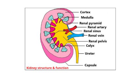
The outer surface of the kidney is convex and the inner surface is concave. The groove in the middle of the kidney is called hilum. The renal artery, renal vein, and ureters are associated with the helium. The kidneys are covered by a layer of connective tissue. This is called the renal capsule (1) & (3).
2. The internal structure of the Kidney
Renal cortex: It is the dark red part on the outside of each human kidney when it is bifurcated vertically is called the renal cortex.
Renal medulla: The light red part on the inside of the kidney is called the renal medulla. About 1 million nephrons are present in the cortex and medulla of each kidney.
Renal pelvis: The funnel-shaped part of the ureters that connect to the concave part of the hilum of the kidney is called the renal pelvis.
Renal pyramid: Parts of the cortex are enlarged in places of the medullary part of the kidney. The enlarged parts of the cortex divide the medulla into several triangular parts, called the renal pyramid.
- Few renal pyramids form the Renal papilla.
- Few renal papillae connected to form Minor calyx.
- Minor calyx combined to form Major calyx.
The renal pelvis is divided into two or three major calyces and four minor calyces. The ureter from the renal pelvis carries the urine from the kidneys directly to the bladder (1) & (3).
The structural and functional unit of the kidney
The nephron is the structural and functional unit of the kidney. It consists of three parts-
- Malpighian corpuscle
- Renal tubule
- Collecting tubule
1. Malpighian corpuscle
It is like a swollen funnel. It is located in the cortex of the kidney. The main parts of the malpighian corpuscle are the bowman’s capsule and glomerulus (1) & (3).
- Bowman’s capsule
This part looks like a cup and is made up of two layers of mantle tissues belonging to the malpighian corpuscle. It acts as a filter and separates the excretory substances from the blood (3).
- Glomerulus
The glomerulus is a cluster of blood vessels from the renal arteries inside the bowman’s capsule. The glomerulus carries blood to the nephron. The diameter of the efferent arteriole of the glomerulus is much less than the diameter of the afferent arteriole. This causes an increase in blood pressure in the glomerulus, which is used for filtration (1) & (3).
2. Renal tubule
The part of the Bowman’s capsule that extends to the collecting tubule is called the renal tubule. The renal tubule consists of three parts (1) & (3).
- Proximal convoluted tubule
The part of the renal tubule extending from the bottom of the Malpighian corpuscle to Henle’s loop is called the proximal convoluted tubule.
- Loop of Henle
Henle’s loop is the part of the renal tubule with a relatively small diameter shaped like the letter ‘U’ below the proximal convoluted tubule. Water is absorbed passively and sodium chloride and potassium ions are absorbed from Henle’s loop. The filtered fluid of the glomerulus is denser in Henle’s loop.
- Distal convoluted tubule
The second spiral tubule of the renal tubule from Henle’s loop is called a distal convoluted tubule. Water is reabsorbed in this part of the renal tubule. From this part, potassium and ammonium ions are secreted and enter the urine inside the tubule.
3. Collecting tubule
The distal convoluted ducts formed by different nephrons that are released into the coarse tubule are called collecting tubules. Many collecting tubules merge to form the Bellini duct, which is finally released in the ureters. After filtration, this duct transports the remaining fluid in the form of urine. Water is absorbed in these tubules and ammonia and potassium ions are secreted (1) & (3).
Role of the kidney in the formation of urine
1. Urine production in the human body
The nephrons in the kidneys play a key role in the formation of urine. Urine is produced as a result of the occurrence of four special processes in the nephron.
2. Ultrafiltration of blood
When the blood mixed with the excretory substance enters the glomerulus, it enters the Bowman’s capsule except for the proteins and blood cells in the plasma. This process is called transfusion, which produces about 160-170 liters of glomerular fluid (or primary urine) per day. It contains nitrogenous excretory substances like water, urea, uric acid, etc., mineral salts, glucose, amino acids, etc. During the flow from the glomerulus to the renal ducts, important elements are absorbed and only 1.5 liters of urine are produced per day till the end.
3. Active reabsorption of ions
Active reabsorption of sodium ions occurs when primary urine enters the Proximal convoluted tubule (PCT) from the glomerulus. The body’s essential substances such as glucose, amino acids, etc. are reabsorbed into the blood through the blood vessels arranged around the ducts. Henley’s loop, distal convoluted tubule, and collecting tubule reabsorbed important components (3).
4. Secretion of excretory substances
Some harmful excretory substances, such as ammonia, benzoic acid, potassium ions, hydrogen ions, and creatine, are secreted from the blood vessels adjacent to the renal tubule. The distal convoluted tubule and the collecting tubule located in the renal ducts participate in the secretion (3).
5. Passive reabsorption of water
With the active absorption of sodium and chloride, water reabsorption occurs in the proximal convoluted tubule of the body. Water is reabsorbed in Henley’s loop and distal convoluted tubule. Water reabsorption occurs in the distal convoluted tubule mainly under the influence of the ADH hormone. Thus, after filtration and reabsorption, thick urine with secretion substances is formed (3).
6. Excretion of urine out of the body
The urine produced in the nephron enters the minor calyx through the Bellini duct, being accepted by the collecting tubule. The urine comes from the minor calyx to the major calyx and eventually reaches the ureters. Urine from the ureters is temporarily stored in the bladder and is excreted out of the body through the urethra (3).
The method of excretion of urine from the nephron is shown in the diagram below
Urine produced in the nephron → collects tubules → Bellini duct → minor calyx → major calyx → ureters → bladder → urethra → excretion outside the body.
Function of kidney
The kidney has many roles in maintaining the balance of the body. Along with excretion, other functions of the kidney are as follows
1. Excretion
The main excretory substance in the human body is urine. Metabolic-contaminated nitrogenous excretory substances are excreted out of the body through the urine prepared in the kidneys. Thus the kidneys help to maintain the internal balance of the body. Urea, uric acid, creatinine, Sulphur, arsenic, etc. are excreted from the body through urine (3).
2. Maintains water balance
The kidneys help maintain water balance in the body and blood by reabsorption. The kidneys reabsorb the essential substances from the purified fluid return them to the blood and keep the blood components normal (1) & (2).
3. Blood pressure control
The kidneys synthesize an enzyme called renin, which plays an important role in maintaining the body’s normal blood pressure (3).
4. Protect the balance of salt
Kidneys maintain the balance of the amount of salt dissolved in the blood.
5. Hormone production
Kidneys produce a hormone called erythropoietin, which helps in the production of red blood cells (RBC) (1) & (2).
