

Apoptosis and necrosis are two types of cell death occurring in the animal body. Faulty cells in the body that can threaten the organism should undergo preplanned self-destruction. Failure to do this will cause cells to proliferate and lead to cancer.
Necrosis is a pathological process detrimental to the body. When cells are exposed to extreme conditions the cell fails to function normally. The cells suffer injury leading to inflammation and tissue damage.
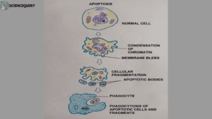
Apoptosis
Definition
It is a pathway of programmed cell death, induced by a tightly regulated intracellular program. Cells that are destined to die activate enzyme caspase, and degrade the cell’s nuclear DNA and cytoplasmic proteins. The cell is phagocytosed. There is no leakage or inflammation.
Apoptosis: It is the way of eliminating unwanted cells or cells damaged beyond repair. Physiological apoptosis causes digit formation in hands and feet during embryogenesis.
Pathological apoptosis is seen during AIDS or cancer.
Steps of apoptosis
Apoptosis takes place through two distinct pathways:
1. Extrinsic pathway
In this pathway, the signal for cell death comes from outside. It is completed in two steps :
A. Initiation phase:
It is initiated through specific receptors called death receptors (surface molecules expressed by cells).
- Fas protein (CD95)
- TNF ( Tumor Necrotic Factor) receptors
- Both these types of death receptors are present on the surface of the cell.
- FAS ligand (Fas L) is a membrane protein expressed mainly on activated T lymphocytes.
- When the T lymphocytes recognize FasL, they bind with the death receptor CD95 or TNF. This ligand is the signal coming from outside so it is extrinsic.
- On the cytoplasmic membrane, they form a domain FADD (Fas-associated death domain).
- FADD now activates pro caspase -8 to active caspase -8.
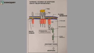
B. Execution phase
It is a convergence point for both the Extrinsic and Intrinsic pathways.
2. Intrinsic pathway
In this pathway, the signal for cell death comes from inside the cell. It is also known as the mitochondrial pathway of apoptosis. It is also completed in two steps:
A. Initiation phase
- Mitochondria contains several proteins capable of inducing apoptosis. A protein cytochrome c is present in between the membranes of the mitochondria.
- On the membrane of mitochondria are also present sensors or antiapoptotic proteins Bcl-2 and Bcl-X. This prevents cytochrome c from coming out from the membranes.
- When the DNA of a cell is damaged it sends a signal to mitochondria to commit suicide. As the signal comes from inside hence it is the intrinsic pathway.
- As mitochondria receive this signal the Bcl-2 and Bcl-X are replaced by proapoptotic proteins. These are Bax, Bak, Bim, and BAD. These allow cytochrome c to come out.
- Cytochrome c enters the cytoplasm and activates caspase 9 by binding with Apaf-1. [Caspase 9 exists in inactive form as it is bound to Apaf-1].
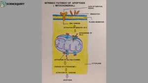
B. Execution phase
- Activated Caspase 8 and 9 will activate two other Caspase – 3 and 7.
- These two caspases now sequentially activate all the caspases in the cytoplasm.
- The cell is now degraded into small apoptotic bodies.
- A phospholipid (phosphatidylserine ) is generally present on the inner surface of the cell membrane. In apoptotic bodies, these phospholipids are flipped to the surface causing it’s tagging.
- Phagocytes recognize these apoptotic bodies and engulf them.
- The process is so efficient that dead cells disappear without leaving a trace. Inflammation is absent.
Necrosis
Cell death due to denaturation of intracellular proteins and enzymatic digestion of lethally injured cells. It is an accidental and uncontrolled form of cell death.
Steps
1. Morphological changes
- Mitochondria swells accompanied by perinuclear localization ( forming a ring around the nucleus). The cristae of mitochondria also show a reduction in number.
- Lysosomes rupture and release their hydrolytic enzymes in the cytoplasm.
- The plasma membrane starts disintegrating and shows myelin figures.
- Increased eosinophilia.
2. Cytological changes
- Overloading of calcium leads to cytotoxic effects especially harmful to neurons.
- Excessive ROS ( reactive oxygen species) production.
- Loss of cytoplasmic RNA.
- ATP levels decrease and a metabolic crisis arises. Kreb’s cycle fails to take place.
- Pyruvate is converted to lactic acid reducing the pH of the cell. Thus cellular enzymes are deactivated.
- Nuclear dissolution due to DNA loss (karyolysis ) is seen. Nuclear fragmentation (Karyorrhexis) also takes place.
3. Other changes
- Proteases like Calpain and Cathepsin activation take place. they cause potential damage to cells. Cyclophilin d also gets activated during necrosis and alters mitochondrial function.
- As cell contents leak, it creates an inflammatory response. This attracts macrophages and dendritic cells to clear off the cell debris.
Types of necrosis
1. Coagulative
Architectural details in a cell persist. Cellular and nuclear details are lost. Hence the cells are called ghost cells. It is the most common type of necrosis.
2. Liquifactive
It is also known as colliquative necrosis. Architectural, cytoplasmic, and nuclear details are lost in this type. So the cells are converted to gel or liquid-like material.
3. Caseous
Here also architectural, cytoplasmic, and nuclear details are lost. It results in a granular, homogenous mass giving the organ a cheese-like appearance. It happens in tuberculosis.
4. Fat necrosis
happens in body organs having excessive fat. It is the death of adipose tissues. Very common in the pancreas and mesentery in the body.
5. Fibrinoid
A special type of necrosis happens in the walls of blood vessels. Injury to tunica media makes deposition of fibrin-like material.
Causes
- Ischemia (lack of blood flow) due to thrombosis or embolism in organs
- Mild burns
- Pus forming bacterial infections.
- Presence of toxins.
Difference between Apoptosis and Necrosis
Basis of differences |
Apoptosis |
Necrosis |
1. Definition |
1. Apoptosis involves the death of a single cell, genetically programmed and energy-driven. | 1.It involves the death of a group of cells, unplanned with a decrease in the ATP reserve of the cell. |
2. Types |
2. Generally physiological, sometimes pathological. | 2. Always pathological. Broadly of two types; coagulative and liquifactive.
Special types like causes, fat, and fibrinoid necrosis are also seen. |
3. Mechanism |
3. Extrinsic and Intrinsic pathway takes place. Each has an initiation phase. The execution phase is a convergence point of both pathway’s initiation phases. | 3. Mechanism varies from organ to organ where necrosis sets in. |
4. Cellular appearance |
4. Cell shrinking and cell membrane is intact | 4. Cell swelling, with disrupted cell membrane. |
5. Nuclear changes |
5. Pyknosis (shrinking)and Karyorrhexis | 5. Karyolysis, pyknosis, and karyorrhexis take place. |
6. Fate of cytoplasm |
6. Cytoplasm is retained in apoptotic bodies. | 6. Cytoplasm is released in the surroundings due to the disintegrating plasma membrane. |
7. Proteins/enzymes involved |
7. Caspases are activated | 7. Calpain, cathepsin and cyclophilin d is activated. |
8. Causes |
8. Due to cell injury and to maintain cellular homeostasis. | 8. Due to ischemia, infection, burns, and the presence of toxins in the body. |
9. Fate of cells |
9. The apoptotic bodies formed are phagocytosed and no cell debris is left. | 9. Due to the lysis of cells, an inflammatory response is set up to clear the cellular debris. |
10. Effect on the body |
10. Beneficial to the body. | 10. It is harmful and can be fatal if left untreated. |
Q&A
1. What is an example of necrosis in humans?
Tuberculosis in lungs.
2. What causes necrosis?
Infection, Injury, diseases, and ischemia are causes of necrosis.
3. What are the four types of cell death?
Apoptosis, Necrosis, Necroptosis, and Pyroptosis.
Summary
- Apoptosis and necrosis are two types of cell death due to injury.
- Apoptosis is programmed cell death. There is no inflammation and cell debris left over from apoptosis.
- Takes place through Intrinsic and Extrinsic pathways. Both pathways have an initiation phase and an execution phase. The execution phase is the convergence point of both pathways’ initiation phase.
- Extrinsic pathway happens when the signal from cell death occurs from outside the cell. The ligand (signal) binds to death receptors(CD95) at the cell’s surface to form the death domain. This death domain activates procaspase 8 to active caspase 9.
- Intrinsic pathway happens when mitochondrial protein cytochrome c leaks outside into the cytoplasm on signal from the nucleus. Cytochrome c activates caspase 9 in the cytoplasm.
- The activated caspase 9 activates caspases 3 and 7 and all other cspases. A plasma membrane phospholipid (phosphatidylserine) is flipped to the outside. This makes phagocytes recognize the apoptotic cell and engulf it.
- Necrosis is cell or tissue death due to irreversible injury like ischemia, burns, toxins, and infections in the body. The cell membrane disintegrates due to lysosomal lysis. Activation of cathepsin in the cytoplasm causes potential damage to the cells. Nuclear dissolution and fragmentation are also seen in necrosis. Inflammatory response and infection are typical of this process.
- Necrosis is mainly coagulative or liquifactive. Special types of necrosis include caseous, fat, fibrinoid, and gangerene. Each has distinct cytological and morphological changes. If left untreated it can lead to severe complications.
Reference
Robbins & Cotran basis of disease by V. Kumar et. al.