Definition
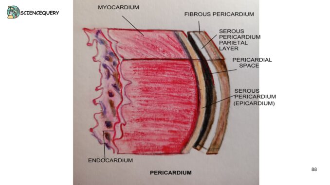
The pericardium is the covering of the heart. It is a fibro-serous (composed of fibrous tissue and serous membrane) sac surrounding the heart and roots of significant blood vessels. It consists of two components :
- Fibrous pericardium
- Serous pericardium
The pericardium lies in the middle of the mediastinum, posterior to the sternum. It is located between the second to sixth costal cartilages. It is situated anteriorly between the fifth to eighth thoracic vertebrae.
Functions
- Prevents over-expansion of the heart when blood volume increases.
- Limits the heart’s movements.
- It serves as a lubricated container in which the heart can contract and relax smoothly.
- Acts as a shock absorber with the help of fluid filled in the pericardial cavity.
Structure
1. Fibrous pericardium
- It is a sac made of tough connective tissue.
- Ihttps://www.amazon.sg/CC-Chatterjees-Human-Physiology-1/dp/B07TFGB31Ms a cone-shaped bag with an apex directed above.
- It defines the boundaries of the middle mediastinum.
- Blood vessels like the ascending aorta, pulmonary arteries, pulmonary veins, and superior and inferior vena cava pierce the fibrous pericardium.
- All of the above blood vessels receive the prolongation of the fibrous pericardium, except the inferior vena cava.
- The fibrous pericardium is attached superiorly to the adventitia of the great vessels and inferiorly to the central tendon of the diaphragm.
- Is also attached anteriorly to the posterior surface of the sternum by ligaments.
- The phrenic nerve also passes through the fibrous pericardium laterally.
- It receives its blood supply from the internal thoracic artery, descending thoracic artery, and musculophrenic arteries.
2. Serous pericardium
- It is thin and consists of the following parts:
- Parietal layer
- Visceral layer/epicardium
- Pericardial cavity
- The parietal and the visceral layers of the serous pericardium are continuous at the root of the great vessels. The narrow space between them is called the pericardial cavity lined by mesothelium.
-
Parietal layer:
- It lines the fibrous pericardium. It is reflected at the root of the blood vessels and becomes continuous as visceral pericardium.
- It develops from the somatopleuric layer of the mesoderm.
- The parietal layer is innervated by the somatic nerve and it’s pain-sensitive.
-
Visceral layer:
- This is closely applied to the heart except at the grooves of the heart. Here it is separated from the heart by blood vessels.
- The visceral layer develops from a splanchnopleuric layer of mesoderm.
- It is innervated by the autonomic nerve and is pain-insensitive.
- Visceral pericardium gets its blood supply from coronary arteries.
- Reflection of the serous pericardium around the large veins forms a recess on the posterior surface of the heart. This is called the oblique sinus. The oblique sinus is closed at one end. It lies behind the left atrium. (Sinuses are passage within the pericardial cavity).
- The transverse sinus is a tunnel-shaped passage. It reflects posterior to the aorta and pulmonary trunk and anterior to the superior vena cava. It lies above and in front of the left atrium.
-
Pericardial cavity:
Is housed between the walls of the serous pericardium. This narrow space is filled by about 50 ml of pericardial fluid. This fluid is secreted by the serous membrane. It reduces friction between membranes during a heartbeat.
Diseases associated with pericardium
Pericarditis
It is an inflammation of the sac. It can be due to virus infection, or from other infections. Medical conditions like heart attack, heart surgery, injuries, and certain medications can cause pericarditis.
Pericardial effusion
The build-up of excess pericardial fluid in the pericardium sac causes this condition. It interferes with the normal pumping action of the heart. Infection from pathogens or rheumatoid arthritis can cause pericardial effusion.
Cardiac Tamponade:
It occurs when fluid or blood collects in the pericardial cavity. This hampers the systole and diastole of the heart. As a result, the body receives insufficient blood. Myocardial infarction, lung cancer, and heart tumors can be the cause of cardiac tamponade.
Constrictive pericarditis
It occurs when the pericardial sac becomes stiff or thicker than normal. This prevents the heart from expanding as it should. This leads to improper filling of blood in the heart leading to heart failure.
Q&A
1. What is the serous pericardium?
It is the membrane inner to the fibrous pericardium. It has two parts-the outer parietal layer and an internal visceral layer enclosing a fluid-filled pericardial cavity.
2. What is the function of the epicardium?
It generates a smooth surface enabling the heart to freely move within the pericardial coelom.
3. What are the three layers of the heart?
The three layers of the heart are the epicardium, myocardium, and endocardium.
4. What is the difference between the epicardium and the pericardium?
Epicardium is the outermost, epithelial layer of the heart.
It is the fibro-serous fluid-filled sac surrounding the muscular body of the heart.
Summary
The pericardium is a fibro-serous sac surrounding the heart and roots of great blood vessels. It consists of two components: Fibrous pericardium and Serous pericardium
- Lies in the middle of the mediastinum, posterior to the sternum.
- It fixes the heart in the mediastinum, limiting its motion. The pericardial fluid between the two layers provides lubrication during contraction and relaxation. Protects the heart from infections.
- A fibrous pericardium is a sac made of tough connective tissue. It is pierced by the great arteries and veins entering and leaving the heart. It is attached to the sternum and diaphragm by ligaments. It is innervated by the phrenic nerve.
- The serous pericardium is thin and has two layers parietal and visceral. Both these layers are continuous with the great vessels. It encloses a fluid-filled pericardial cavity, lined by mesothelium.
- Diseases of the pericardium include pericarditis, cardiac tamponade, and pericardial effusion.
Reference
C. C. Chatterjee’s Human Physiology: Volume 1.
Written By: Ahana Mitra
